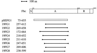
FIG. 2.
Schematic presentation of the Fbe protein and alignment of inserts from nine phagemid clones obtained after panning against Fg. The different regions are indicated by S (the signal sequence), A (the Fg-binding region), and R (the highly repetitive region). The insert of the single clone (pSEFG1) originated from strain HB is shown as an open bar, while the eight clones derived from strain 19 are presented by solid lines. The numbers indicate the positions of amino acids (aa) in the Fbe protein as defined in the legend to Fig. 4.

