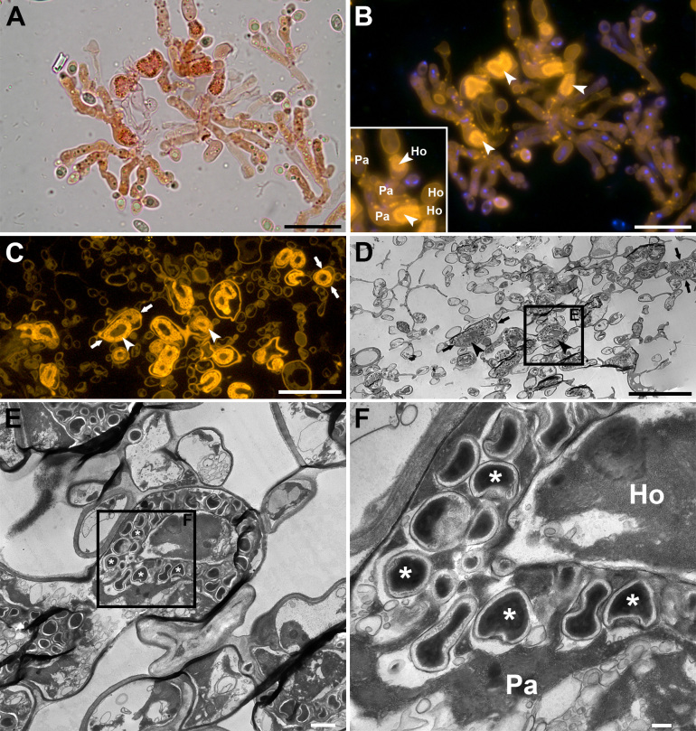
Fig. 2 .
Brightfield, epifluorescence and transmission electron microscopy (TEM) of Colacogloea Universitatis-gandavensis sp. nov. A, B. Whole-mount preparation, stained with Congo red and DAPI, visualised using brightfield (A) and epifluorescence (B) microscopy. Epifluorescence microscopy facilitates fast detection of colacosomes as they exhibit bright fluorescence signals. Inset shows the intricate host–parasite (Ho-Pa) interface. Arrowheads indicate regions of colacosome clustering. Note the occurrence of individual colacosomes in parasite tissue (bright spots). C, D. Serial sections of a Spurr-embedded sample, showing the same region. Corresponding structures are indicated with arrows. (C) Section stained with Congo red and visualised using epifluorescence microscopy. (D) Equivalent serial section of the same region as in (C), visualised using TEM. E, F. High-magnification details of colacosome clusters (arrowheads), composed of many individual colacosomes (asterisks), arranged in parasitic hyphae (Pa) along the host–parasite interface (Ho-Pa), showing their typical electron dense cores. Scale bars: A–D = 20 μm, E = 10 μm, F = 200 nm.

