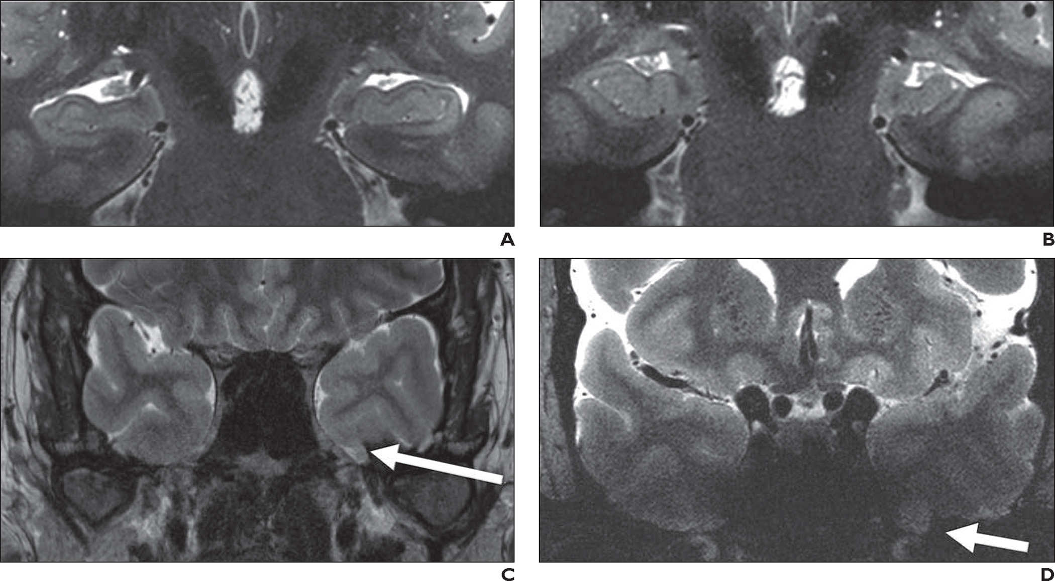
Fig. 10—

Epilepsy and hippocampal imaging.
A, 48-year-old healthy man who volunteered to undergo 7-T brain MRI. Oblique coronal T2-weighted turbo spin-echo (TSE) image (slice thickness, 1.5 mm; FOV, 0.2 × 0.2 mm) shows excellent anatomic detail of hippocampus bilaterally.
B, 35-year-old man with complex partial seizures for 13 years and left temporal spike-wave complexes on electroencephalography (EEG). Oblique coronal 7-T T2-weighted TSE image obtained with same parameters and at comparable slice position as A shows small left hippocampus with loss of interdigitations in comparison with contralateral normal side, consistent with mesial temporal sclerosis.
C and D, 35-year-old man with complex partial seizures from left temporal lobe per EEG (different patient from B). Oblique coronal 3-T T2-weighted image (C) shows left encephalocele (arrow, C), which was suspected cause of seizures. Oblique coronal 7-T T2-weighted TSE image (D) was unable to clearly show left temporal encephalocele despite use of dielectric pads and neck extension; encephalocele (arrow, D) was identified only after retrospective review of prior 3-T MRI (C).
