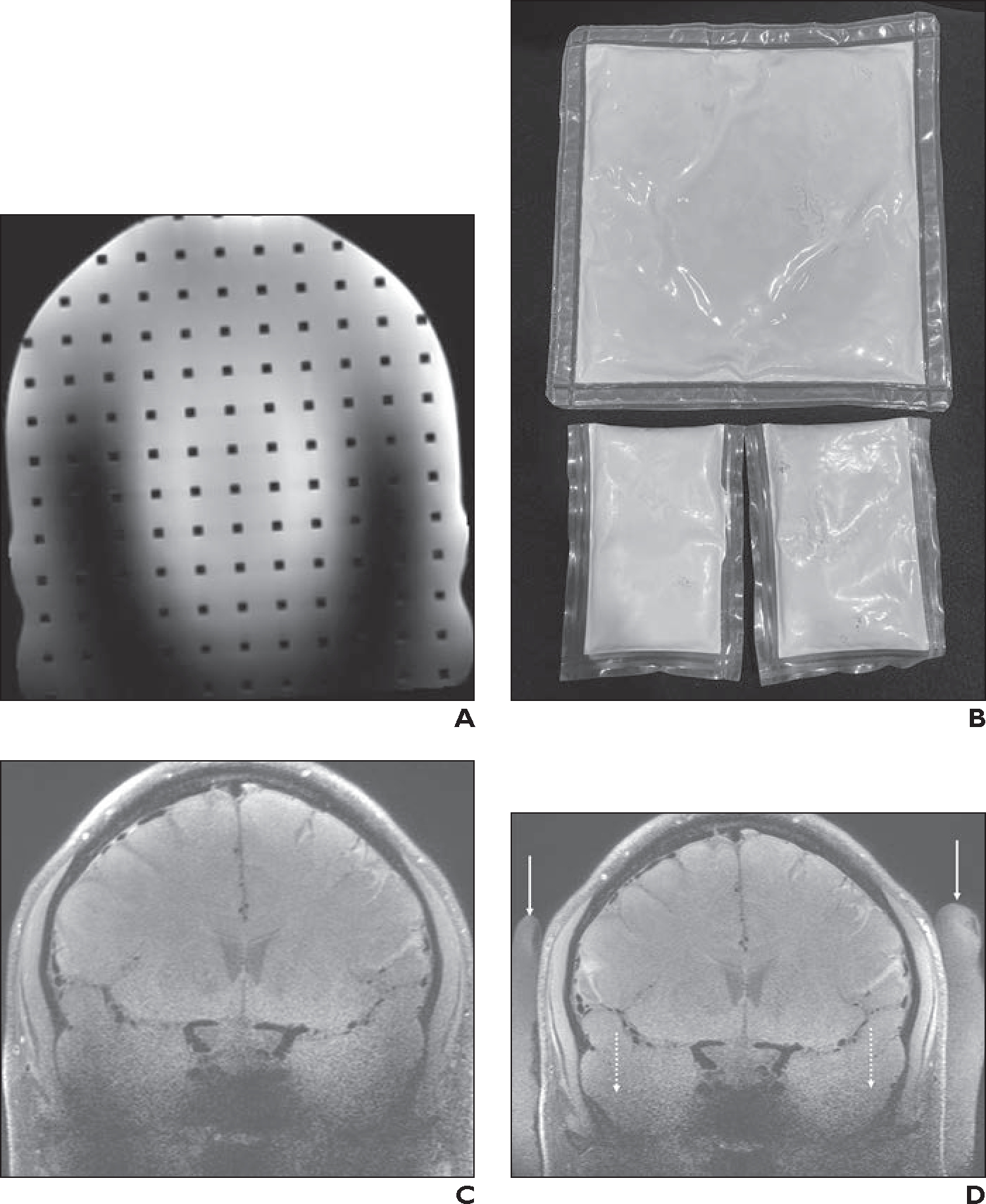
Fig. 11—

Transmit B1 inhomogeneity artifacts.
A, Coronal 3D 7-T T2-weighted SPACE MR image of head phantom obtained with 1-channel transmit/32-channel receive array head coil shows U-shaped region of poor signal in z-axis caused by B1 inhomogeneity artifacts.
B, Photograph shows dielectric pads. Because of its large size, commercially available dielectric pad (top) typically can be placed only in suboccipital region. Smaller dielectric pads made in house (bottom) are easier to place in certain areas, such as temporal region.
C and D, 40-year-old healthy man who volunteered to undergo 7-T brain MRI. Coronal 3D T1-weighted SPACE images were obtained at level of carotid terminus with black-blood vessel wall imaging sequence without (C) and with (D) dielectric pads placed along right and left lateral aspects of head. Whereas commercially available pad is not visible on MRI, in-house dielectric pads (solid arrows, D) are. Use of dielectric pads markedly improves SNR of parenchyma near lower skull base (dotted arrows, D) and improves delineation of bony structures, secondary to decreased B1 artifacts. However, hypointense signal near sphenoid sinuses persists after placement of dielectric pads.
