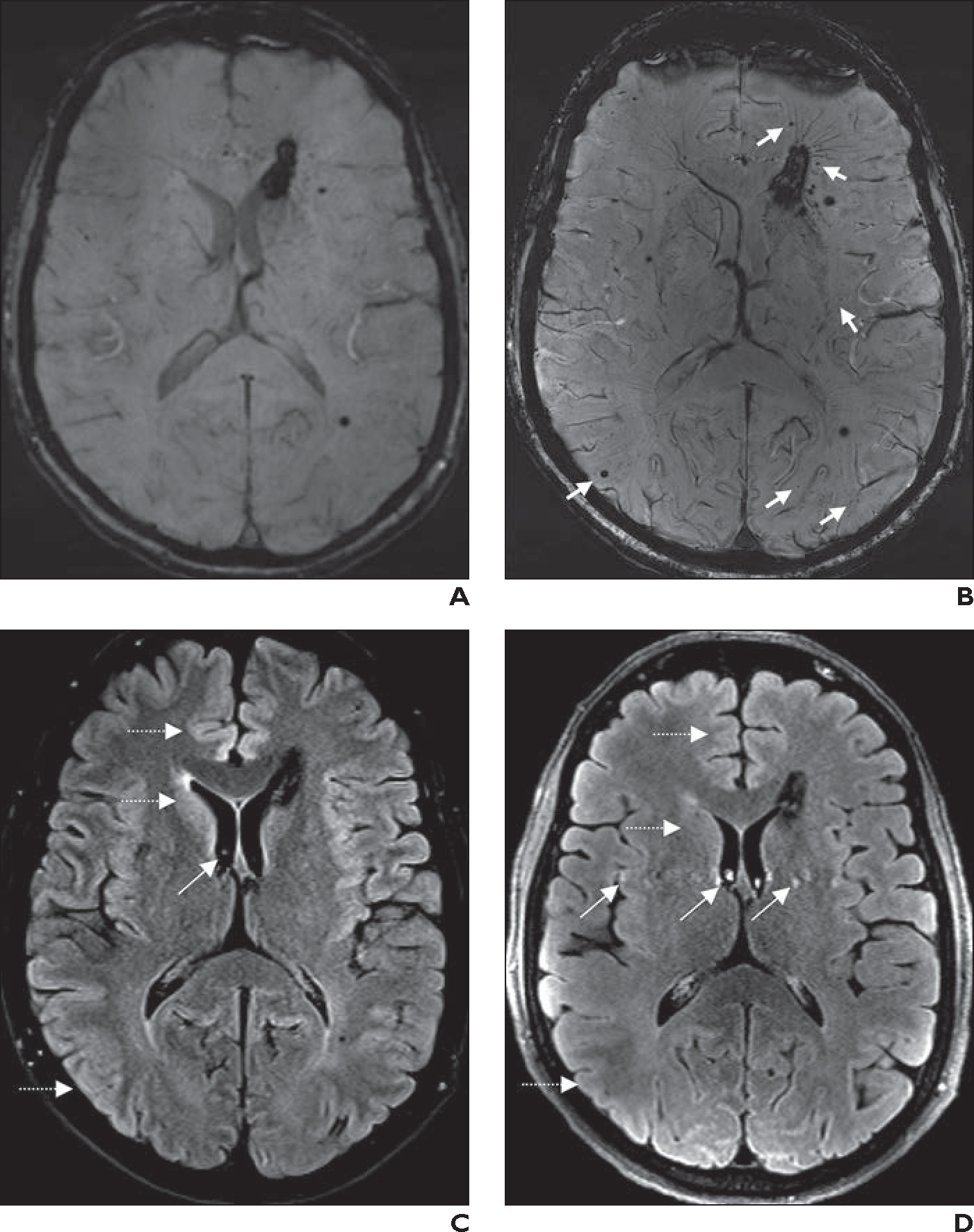
Fig. 2—

56-year-old man with known multiple cerebral cavernomas.
A, Axial 3-T susceptibility-weighted imaging (SWI) shows cavernomas, which are stable compared with appearance on prior surveillance MRI examinations performed over many years.
B, Axial 7-T SWI obtained 18 months after A shows additional tiny cavernomas (arrows), which were not visible at 3 T, and greater conspicuity of draining veins associated with largest periventricular cavernoma.
C, Axial 2D 3-T fat-saturated FLAIR image from same examination as A shows subtle CSF pulsation artifacts (oblique arrow). Gray-white matter differentiation (horizontal arrows) in frontal lobes, basal ganglia, temporal lobes, and insular cortexes is better than at 7 T.
D, Axial 2D 7-T fat-saturated FLAIR image from same examination as B shows more pronounced CSF pulsation artifacts (oblique arrows). Gray-white matter differentiation (horizontal arrows) in frontal lobes, basal ganglia, temporal lobes, and insular cortexes is worse than at 3 T.
