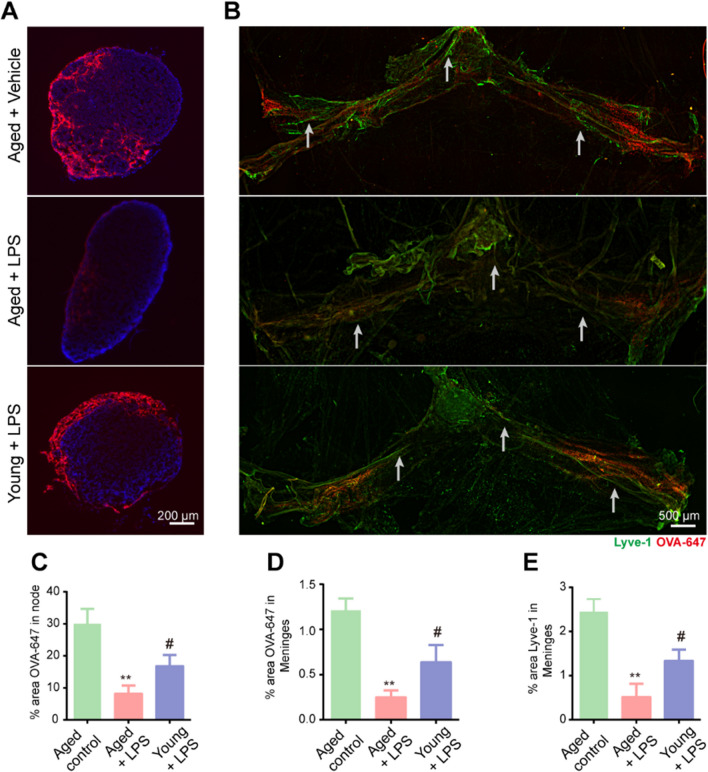
Fig. 3.
Meningeal lymphatic function in aged mice is more vulnerable to sepsis. A Representative immunofluorescence images of OVA-647 accumulation in dCLNs. B Representative immunofluorescence images of meningeal whole-mounts of OVA-647 stained with DAPI and Lyve-1. C Graph depicting the percentage area of OVA-647 coverage in the dCLNs. D Graph depicting the percentage area of OVA-647 coverage in the meningeal lymphatic vasculature. E Graph depicting the percentage area of Lyve-1 coverage in the meningeal lymphatic vasculature. n = 5. **P < 0.01, aged control group vs. aged + LPS group, # P < 0.05, young + LPS group vs. aged + LPS group. All data are expressed as the mean ± SD

