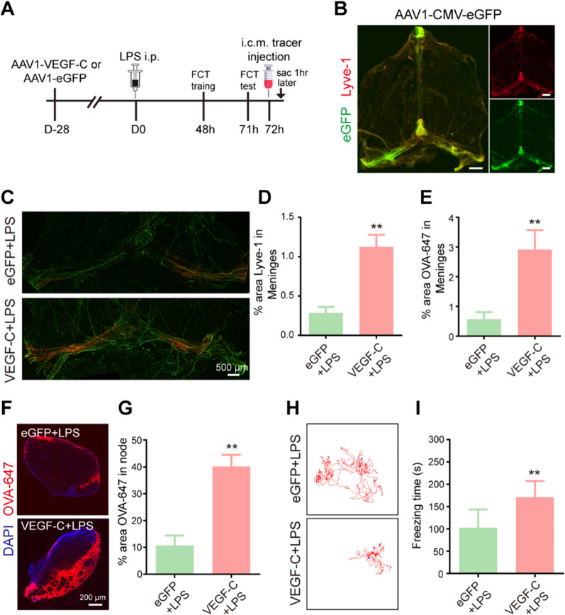
Fig. 4.
Improvement of meningeal lymphatics alleviates sepsis-induced cognitive dysfunction in aged mice. A The experimental timeline of the intracisternal AAV infusion, LPS treatment, behavioral test, tracer injection into the cisterna magna, and tissue collection (sac). B Representative images depicting eGFP (green)-labeled AAV1-infected cells surrounding Lyve1 (red)+ meningeal lymphatics, scale bars: 1 mm. C Representative immunofluorescence images of meningeal whole-mounts of OVA-647 stained with Lyve-1. D Graph depicting the percentage area of Lyve-1 coverage in the meningeal lymphatic vasculature, n = 6. E Graph depicting the percentage area of OVA-647 coverage in the meningeal lymphatic vasculature, n = 6. F Representative immunofluorescent images of OVA-647 accumulation in dCLNs. G Graph depicting the percentage area of OVA-647 coverage in the dCLNs, n = 6. H Representative trajectory of each group in the FCT. I Quantitative of freezing time in the FCT, n = 8. **P < 0.01. All data are expressed as the mean ± SD

