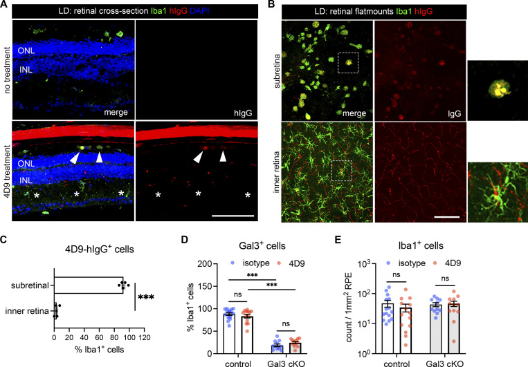
Figure S4.
Subretinal microglia with 4D9 treatment. (A) Staining of human IgG (red) and Iba1 (green) in retinal cross-sections collected from mice with or without 4D9 treatment in LD. The hIgG is used to trace 4D9 antibodies, which outlines retinal vasculatures in 4D9-treated mice. Arrowheads indicate the presence of 4D9 antibodies in the subretinal microglia, while asterisks indicate the absence of 4D9 antibodies in microglia from the inner retina. INL, inner nuclear layer. (B) Human IgG (red) and Iba1 (green) staining in RPE and neuroretina flatmounts were collected from mice treated with 4D9 antibodies in LD. (C) Quantifications of hIgG+ microglia in the subretinal space and neuroretina. (D and E) Quantifications of Iba1+ cells and Gal3+ cells between control and Gal3 cKO mice treated with either isotype or 4D9 (n = 13 per group). Scale bars: 100 μm. Data were collected from two to four independent experiments. ***: P < 0.001; ns: not significant. Unpaired Student’s t test (C); two-way ANOVA with Tukey’s post hoc test (D and E).

