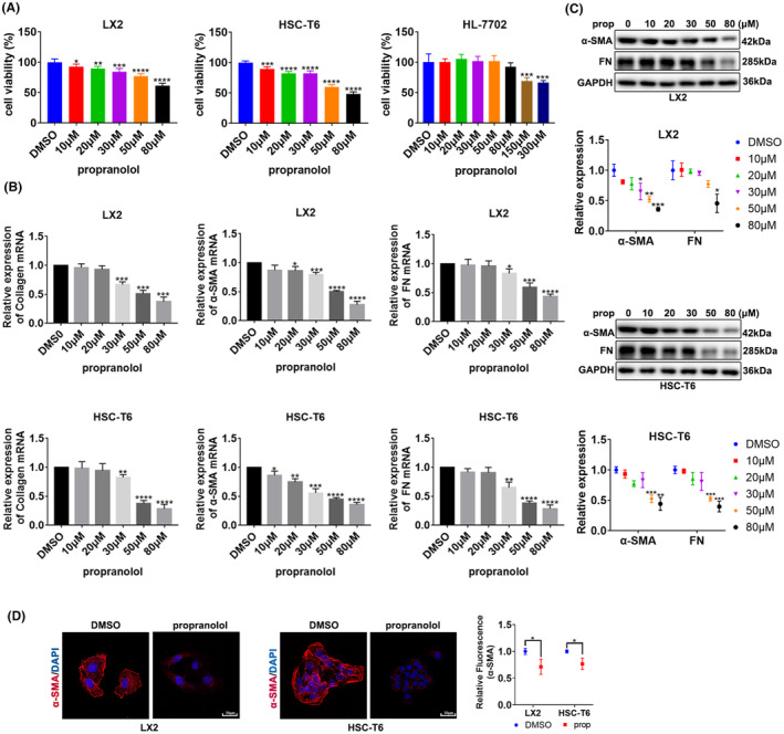
FIGURE 1.

Propranolol inhibits the viability and activation of HSCs. (A) Viability of HSC cell lines LX2 and T6 and normal liver cell line HL‐7702 treated with different concentrations of propranolol. (B, C). LX2 and T6 cells were treated with different concentrations of propranolol or DMSO for 24 h, and the levels of α‐SMA, collagen and fibronectin (FN) were determined. (D) LX2 cells was treated with 80 μM propranolol or DMSO, and T6 cells were treated with 50 μM propranolol or DMSO; α‐SMA were revealed by immunofluorescence. Data are presented as mean ± standard error from three independent experiments (*p < 0.05, **p < 0.01, ***p < 0.001, ****p < 0.001).
