
Fig. 1 |. Multimodal speech decoding in a participant with vocal-tract paralysis.
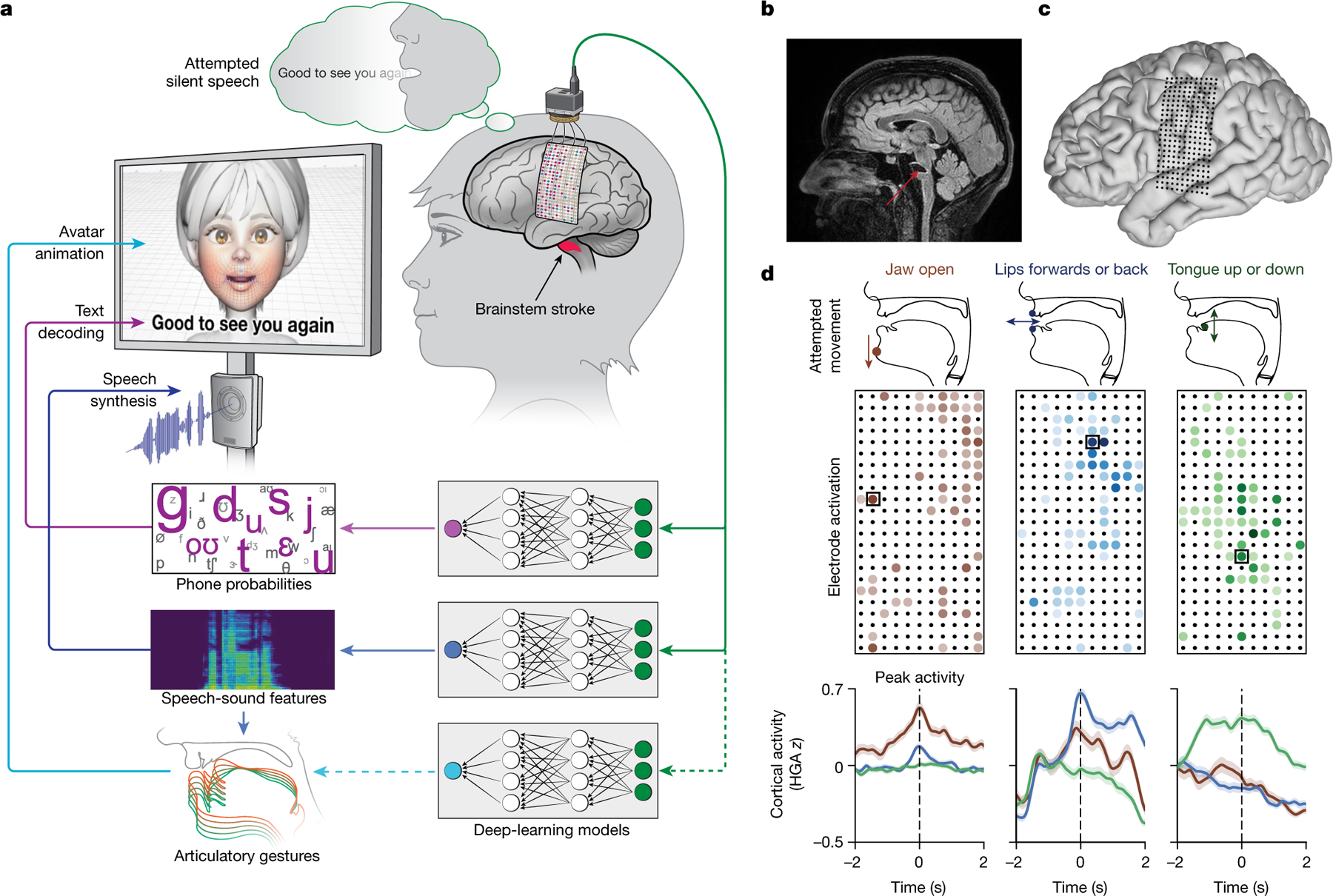
a, Overview of the speech-decoding pipeline. A brainstem-stroke survivor with anarthria was implanted with a 253-channel high-density ECoG array 18 years after injury. Neural activity was processed and used to train deep-learning models to predict phone probabilities, speech-sound features and articulatory gestures. These outputs were used to decode text, synthesize audible speech and animate a virtual avatar, respectively. b, A sagittal magnetic resonance imaging scan showing brainstem atrophy (in the bilateral pons; red arrow) resulting from stroke. c, Magnetic resonance imaging reconstruction of the participant’s brain overlaid with the locations of implanted electrodes. The ECoG array was implanted over the participant’s lateral cortex, centred on the central sulcus. d, Top: simple articulatory movements attempted by the participant. Middle: Electrode-activation maps demonstrating robust electrode tunings across articulators during attempted movements. Only the electrodes with the strongest responses (top 20%) are shown for each movement type. Colour indicates the magnitude of the average evoked HGA response with each type of movement. Bottom: z-scored trial-averaged evoked HGA responses with each movement type for each of the outlined electrodes in the electrode-activation maps. In each plot, each response trace shows mean ± standard error across trials and is aligned to the peak-activation time (n = 130 trials for jaw open, n = 260 trials each for lips forwards or back and tongue up or down).