Figure.

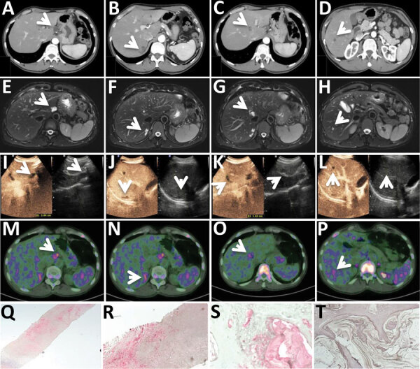
Diagnostic tests for patient in Italy with confirmed autochthonous case of human alveolar echinococcosis, 2023; white arrows indicate lesions. A–D) Contrast-enhanced computed tomography arterial phase. E–H) T2-weighted magnetic resonance imaging. I–L) Ultrasonography and contrast-enhanced ultrasonography. M–P) 18F-FDG-PET scan delayed acquisition (4 hours). Q–T) Em2 immunohistochemistry indicating small particles of Echinococcus multilocularis (spems) stained in red in patient’s sample (original magnification ×2.5 [Q] and ×20 [R]); positive alveolar echinococcosis sample control (original magnification ×20 [S]); Em2 negative control (cystic echinococcosis case, negative laminated layer; original magnification ×20) (T).
