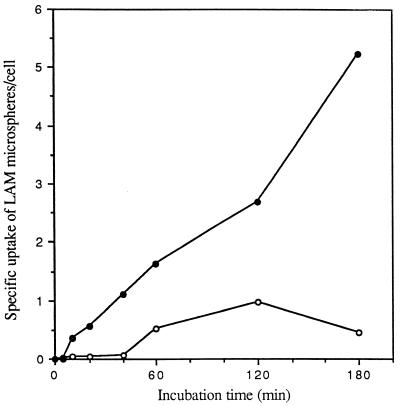
FIG. 1.
Specificity of cell association of LAM microspheres with monocytes and MDMs. Monocytes and MDMs were incubated with 2 × 10⁷ LAM- or HSA (control)-coated microspheres for the indicated time at 37°C. At the end of each incubation period, monocytes and MDMs were fixed with formalin. The total number of LAM or HSA microspheres associated with the cell was enumerated by phase microscopy. The specific uptake of LAM microspheres by monocytes (open circles) and MDMs (solid circles) was obtained by subtracting the total number of associated HSA microspheres from that of LAM microspheres at each point for each cell type. All points are means from two independent experiments.

