
Figure 1.
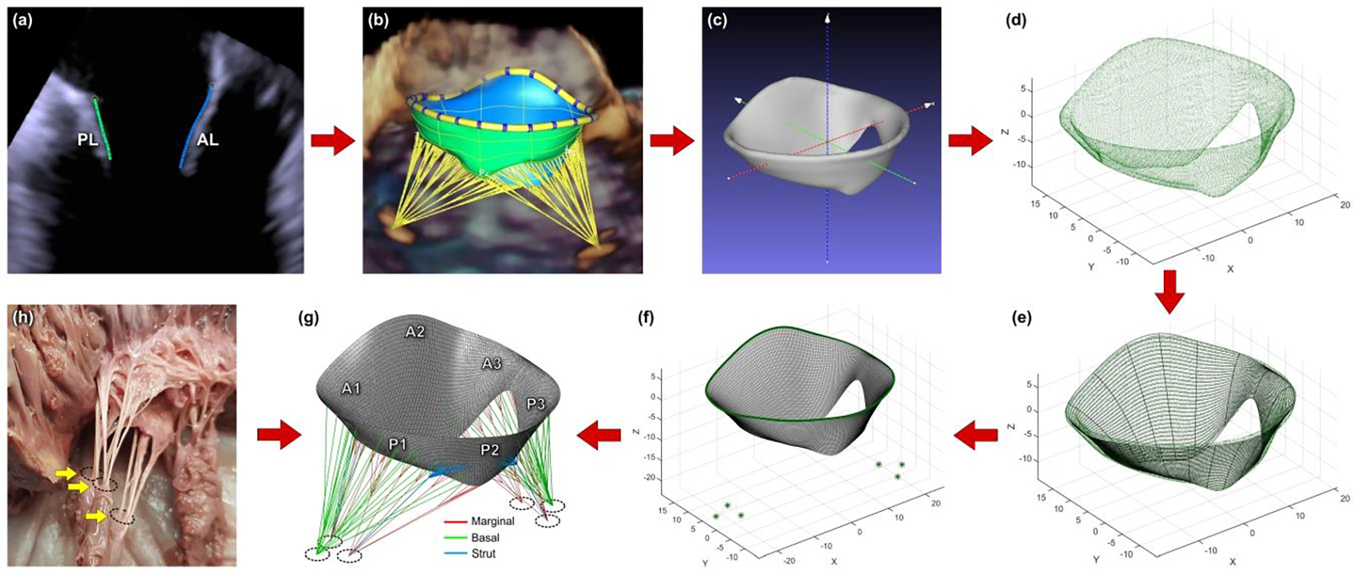
Steps for creating a patient-specific mitral valve computational model: (a) MV leaflets segmented in Siemens AutoValve; (b) complete geometry of the valve ready to be exported as STL model; (c) model positioned in the 3D Cartesian coordinate system; (d) model imported into MATLAB as a point cloud; e) mid-surface of the leaflets approximated with splines; (f) mesh created using triangular shell elements; (g) finished finite element model with chordae tendineae; (h) ex vivo mitral valve specimen showing chordae emerging from three papillary muscle heads.