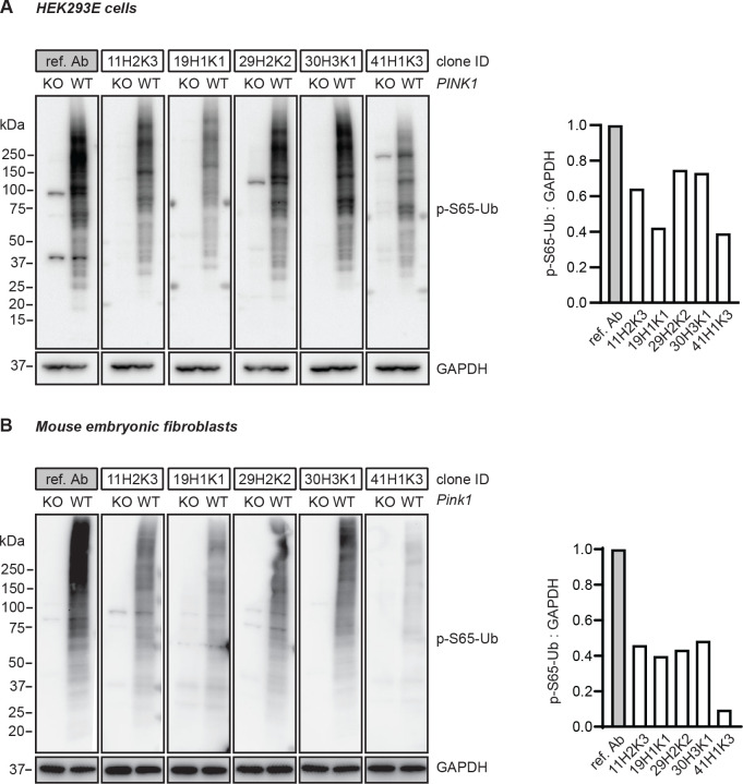
Figure 2.
Western blot assessment of the top p-S65-Ub antibodies in cells. (A) HEK293E cells and (B) mouse embryonic fibroblasts with (WT) or without PINK1 expression (PINK1 KO or pink1 KO) were treated with mitochondrial depolarizer and analyzed by western blot. Representative western blot images are shown for the performance of the top five p-S65-Ub antibody clones in each cell type. Note that concentrations of the tested p-S65-Ub antibodies are five times higher compared to the reference antibody. GAPDH was used as loading control. A densitometric quantification of p-S65-Ub relative to GAPDH is shown alongside after normalization by the reference antibody set to 1. Ref. Ab - reference antibody, KO - knockout, WT - wild-type.

