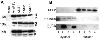
FIG. 3.
Viral protein expression in A549 cells infected with wt rRSV and the NS1/NS2 gene deletion mutants. (A) Wt rRSV and the NS1/NS2 deletion mutants display comparable levels of gene expression, as illustrated by the levels of N and P proteins. A549 cells were infected with the indicated viruses at an MOI of 3 PFU/cell and harvested 6 h, 10 h, or 14 h postinfection. Equivalent amounts of the cell lysates were subjected to SDS-PAGE and Western blot analysis with a polyclonal serum raised against purified RSV virions. The portion of the blot containing the N and P proteins is shown. (B) NS1 and NS2 are located predominantly in the cytosolic fraction of wt-rRSV-infected A549 cells. Cells were either mock infected (lanes 1) or infected with wt rRSV (lanes 2), ΔNS1 (lanes 3), or ΔNS2 (lanes 4) at an MOI of 3. The cells were harvested 20 h postinfection, and nuclear and cytosolic fractions were prepared and analyzed by Western blotting. The nuclear fraction is represented in fourfold-greater relative concentration than the cytosolic fraction. The efficacy of cell fractionation was confirmed using antibodies to USF2 as a marker for nuclear content and α-tubulin as a marker for cytosolic content. The localization of NS1 and NS2 was identified using a polyclonal antiserum that reacts with both proteins but with NS1 more efficiently than NS2.

