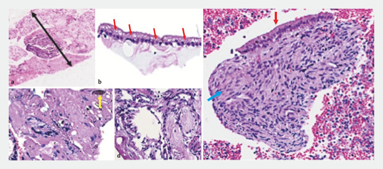
Fig. 2.
Ultrasound-guided biopsies appearance of serous cystic neoplasia a ProCore 20-gauge caliber of the specimen (black arrow with two heads). b Simple cubic epithelium with vacuoles containing glycogen (red arrows). c Dense cicatricial stroma with hemosiderin (yellow arrow). d Loose myxoid stroma. e Mucinous cystic neoplasm. Flat epithelium with mucoproducing gastric foveolar pattern (red arrow) and dense ovarian pattern stroma (blue arrow).

