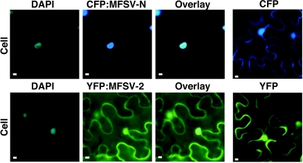
FIG. 6.
Epifluorescence microgaphs of subcellular localizations of fluorescent-protein fusions of the MFSV N and ORF2 proteins. The DNA selective dye DAPI was used to determine the positions of nuclei in plant cells. Infiltrations of unfused CFP and YFP were included as negative controls. Cellular views of localizations of CFP-MFSV N to the nucleus (top row), and YFP-MFSV 2 throughout the cell (bottom row) are shown. Bars = 5 μm.

