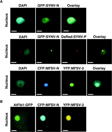
FIG. 8.
Subcellular localization of fluorescent-protein fusions of the MFSV N and ORF2 proteins and the SYNV N and P proteins. The DNA selective dye DAPI was used to determine the positions of nuclei in plant cells. (A) Epifluorescence microgaphs of GFP-SYNV N infiltrated by itself targeting the nucleus (top row); coinfiltrated GFP-SYNV N and DsRed-SYNV P targeting the subnuclear locale (middle row) (see also Goodin et al. [13]); and coinfiltrated CFP-MFSV N and YFP-MFSV 2 targeting the nucleolus (bottom row). (B) Confocal micrographs of the colocalization of AtFib1-GFP, CFP-MFSV N, and YFP-MFSV 2 in the nucleolus of a plant cell. Single-plane optical sections (0.3 mm thick) for each channel were taken through the largest area of fluorescence within the nucleolus. Bars = 5 μm.

