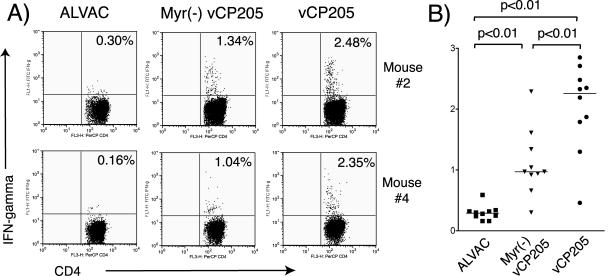
FIG. 6.
HIV-specific CD4+ T-cell responses measured by intracellular-cytokine staining. Splenocytes from naïve BALB/c mice were infected with vaccinia virus expressing Env and Gag to function as APCs. Cryopreserved splenocytes from immunized mice were restimulated with APCs at the ratio of 4 to 1 for 7 h. The splenocytes were then subjected to surface labeling for CD4 and intracellular cytokine staining for IFN-γ. (A) Results from two representative mice from groups immunized with ALVAC, Myr− vCP205, and vCP205. (B) Cumulative results from the three groups of mice. The horizontal lines represent the mean responses for each group.

