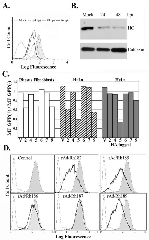
FIG. 4.
MHC I expression in RhCMV-infected cells and in the presence of individual RhCMV US6 family proteins. (A) Flow cytometry of RhCMV-infected fibroblasts. TRFs were infected with RhCMV (MOI, 2) for the indicated times (hpi, hours postinfection), and surface levels were monitored with W6/32 antibody. (B) Immunoblot of MHC I and calnexin using cell lysates from TRFs infected with RhCMV (MOI, 2) for the indicated times. MHC I was detected with antiserum K455. Anti-calnexin antiserum was obtained from Stressgen. (C) RhCMV US6 family genes, either wild type or HA tagged, were cotransfected with a GFP expression vector into either HeLa cells or TRFs. MHC I surface expression was monitored by flow cytometry using the monoclonal antibody W6/32 at 42 h posttransfection. Transfection efficiencies, determined by GFP fluorescence, were ∼80 and 30% for HeLa cells and TRFs, respectively. The bars in the graph represent the ratio of the mean W6/32 fluorescence (MF) of GFP-positive cells to that of GFP-negative cells. The numbers (2, 4, 5, 6, 7, and 9) on the x axis represent Rh182, Rh184, Rh185, Rh186, Rh187, and Rh189, respectively, and V is the vector control. (D) MHC I surface expression upon transduction with adenovirus constructs. HeLa cells were infected with recombinant adenovirus (MOI, 25) for 36 h. As a control, cells were infected with tet-transactivator-expressing adenovirus (rtetA) (shaded). The cells were either stained with W6/32 (solid line) or unstained (broken line). Note the reduction of surface levels in cells transduced with rAd/Rh182, rAd/185, or rAd/Rh189.

