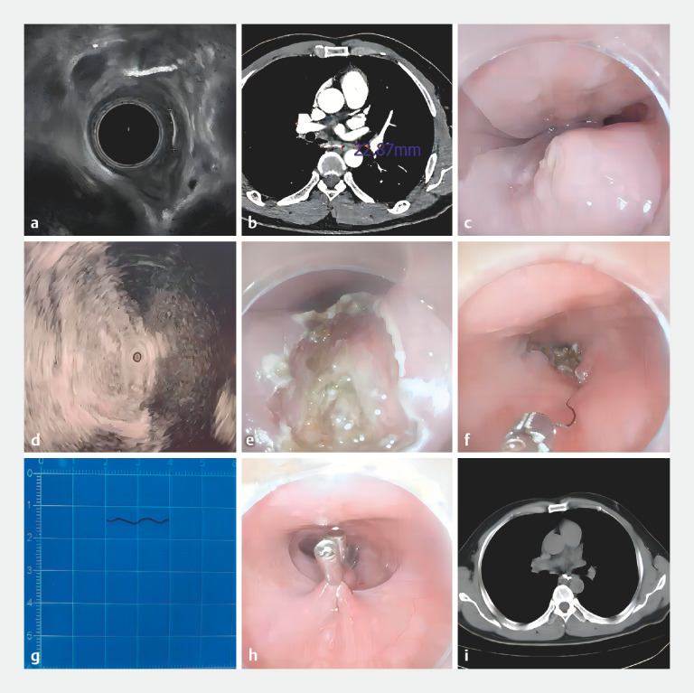
Fig. 1.
Illustration of the major steps involved in endoscopic removal of an esophageal foreign body embedded in the muscularis propria. Endoscopic removal of the entirely embedded esophagus-penetrating foreign body in a 53-year-old man presenting with a 3-week history of retrosternal pain. a Visualization of the foreign body under endoscopic ultrasonography. b Visualization of the foreign body under enhanced computed tomography. c Visualization of the granulation tissue protruding from the posterior esophageal mucosa, with a small amount of purulent discharge. d Localization of the foreign body with the guide of the ultrasound probe. e,f Myotomy, foreign body exposure, and extraction. g The foreign body was a curved metal wire, 20 mm in length. h Closure of the esophageal wound. i Postoperative computed tomography of the chest showed no residual foreign body left.

