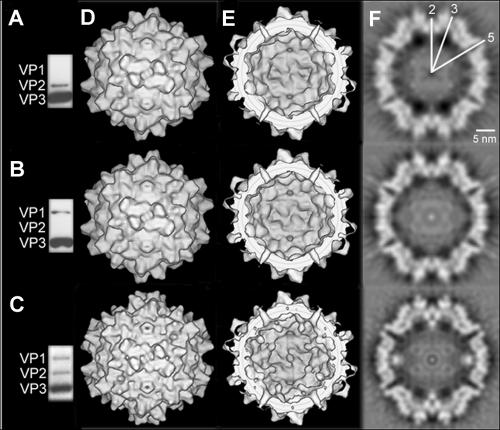
FIG. 1.
Western blot analysis and 3D image reconstruction of empty ΔVP1, ΔVP2, and wild-type capsids. The presence of the VP proteins in wild-type and mutant empty capsids was determined by Western blot analysis using the antibody B1, which detects the C termini of all three VP proteins. The ΔVP1 capsids (A), the capsids devoid of VP2 (B), and wild-type empty capsids (C) are shown. The outer surfaces of the 3D image reconstructions of ΔVP1- (top), ΔVP2- (middle), and wild-type particles (bottom) are viewed down a twofold symmetry axis (D), as well as the inner surfaces (E). Equatorial slices of the reconstructions are also shown (F). Some of the symmetry axes are marked in white.

