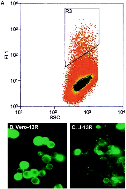
FIG. 2.
Fluorescence-activated cell sorting of J-13R cells and surface fluorescence of J-13R and Vero-13R cells. Panel A. G418-resistant cells in 75-cm2 flasks were removed and surface stained by FITC-conjugated anti-IL13Rα2 antibody as described in Materials and Methods. Flow cytometry was performed on a FACScalibur (Becton-Dickinson, Mountain View, CA). Panels B and C represent FITC-positive Vero-13R and J-13R cells, respectively. The FITC-positive cells, approximately 5 × 10⁴ cells, comprising 3 to 5% of the total cell population, were collected and amplified.

