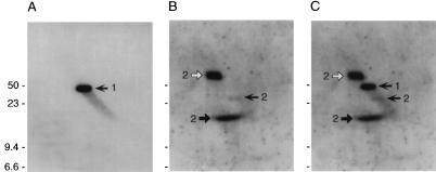
FIG. 4.
Southern blot analysis of B. burgdorferi N40 plasmids separated by two-dimensional agarose gel electrophoresis. (A) Hybridization with an N40-dbpA probe. The single DNA species is indicated by a left-facing arrow and a numeral 1. (B) Hybridization with a probe derived from a gene located immediately 3′ of the p21 gene, a gene located on a 32-kb circular plasmid. This probe hybridized with three DNA species, each labeled with a numeral 2: supercoiled circular plasmid (solid right-facing arrow); nicked, open circular plasmid (open right-facing arrow); and linearized plasmid (solid left-facing arrow). (C) Alignment of the blots shown in panels A and B. Note that the linear and open circular DNAs migrate on one axis, while supercoiled DNA migrates on a different axis. Sizes are indicated in kilobases.

