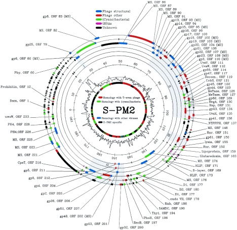
FIG. 1.
Organization of the genome of phage S-PM2. The circles from outside to inside indicate the following: 1 to 6 represent the six reading frames, 7 is the scale bar (in kilobases), 8 is G+C content (smoothed with a sigma = 200 bp; Gaussian; range is 28.2 to 60.4%, with a mean of 37.8%), and 9 shows homology with other organisms. Labels show ORF numbers and gene designations where a putative homologue in T4 has been identified. MS indicates that presence of protein in the virion has been established by mass spectrometry and is shown in parentheses if the ORF has already been shown to encode a virion structural protein on the basis of proposed homology. The following color scheme has been used: green, ORFs encoding proteins exhibiting similarity to cyanobacterial proteins; blue, phage structural proteins; red, other phage proteins; purple, tRNA genes; and black, unidentified ORFs. The four clusters of T4-like genes are shaded in gray.

