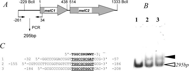
FIG. 5.
(A and B) Gel mobility shift assay (B) for detection of the binding of AdpAl to a γ-32P-labeled 295-bp DNA fragment (A) covering the promoter region of the melC operon. The solid arrowhead in panel B indicates the position when 0.01 μg (lane 2) or 0.02 μg (lane 3) of total proteins isolated from E. coli BL21(DE3) carrying pJTU1464 was added to the radioactively labeled probe fragment (open arrowhead). Addition of 0.02 μg of total proteins isolated from E. coli BL21(DE3) carrying pET15b to the same probe was used as a negative control (lane 1). (C) Alignment of the three regions upstream of the melC operon (A) with the consensus AdpA-binding sequence (51) shown at the top (5′-TGGCSNGWWY-3′, where S is G or C, W is A or T, Y is T or C, and N is any nucleotide).

