Fig. 3.
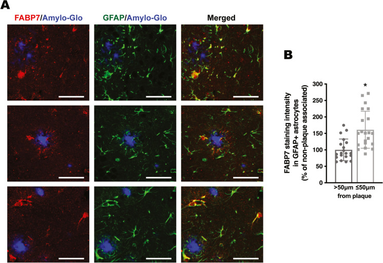
FABP7 immunostaining in astrocytes located in the vicinity of amyloid plaques. A Representative microphotographs showing FABP7 immunoreactive astrocytes surrounding amyloid plaques in the cerebral cortex of 9-month-old APP/PS1 mice. Amyloid plaques were stained using Amylo-Glo (blue), followed by co-immunostaining with antibodies specific for FABP7 (red) and GFAP (green). Scale bar: 50 μm. B Quantification of FABP7 staining intensity in GFAP+ astrocytes located within 50 μm from the edge of a plaque (plaque-associated) or beyond this boundary (non-plaque-associated; > 50 μm from the edge of a plaque). Data are expressed as a percentage of non-plaque-associated GFAP+ astrocytes. 2–4 images per animal were analyzed, n = 4–5 animals/genotype; data are expressed as mean ± SD, *p < 0.05

