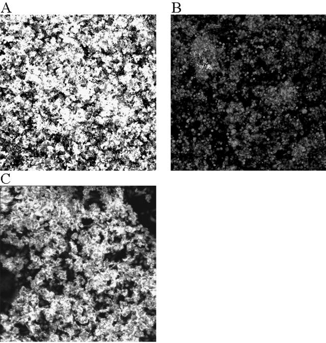
FIG. 1.
Comparison of biofilm formation by serovar Typhimurium strain BJ2710 or serovar Typhimurium strain BJ2508 (BJ2710 ΔfimH) on cultured HEp-2 cells. The composite image of biofilm formed by adherent serovar Typhimurium strain BJ2710 (A) or nonadherent BJ2508 (BJ2710 fimH) (B) was recorded after 24 h of incubation in the biofilm flowthrough system. (C) Biofilm formed by the fimH mutant strain BJ2508 complemented with pISF204, which carries an intact LT2 fimH gene. Panel A demonstrates that extensive biofilm is formed by strain BJ2710. Mutation of the fimH gene (B) abrogates the ability to form biofilm. The bacteria were labeled with GFP and appeared green in the confocal microscope, and the HEp-2 cells were stained with CMTMR and appeared red in the confocal microscope. The level of cell stain for each of the samples was comparable to what is observed in panel B, which is almost exclusively HEp-2 cell staining.

