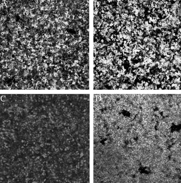
FIG. 6.
Comparison of biofilm formation by serovar Typhimurium strain BJ2710 and serovar Typhimurium BJ3458 (BJ2710 hns::cam) on cultured HEp-2 cells. Composite images of biofilm formed by adherent serovar Typhimurium strain BJ2710 (A) or nonadherent BJ2508 (BJ2710 fimH) (C) were recorded after 24 h of incubation. Panel B is an image of biofilm formed by BJ2710 hns (BJ3458), and panel D is biofilm formed by BJ2710 hns (BJ3458) complemented with plasmid pBDJ254, which carries an intact hns gene. Absence of the hns gene has little effect on biofilm formation (B), but overexpression of the hns gene resulted in a reduced ability to form biofilm (D). The bacteria carried GFP and appeared green with the confocal microscope, and the HEp-2 cells were labeled with CMTMR and appeared red with the microscope.

