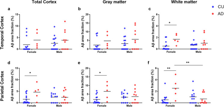
Fig. 3.
Sex-specific differences in the amyloid-beta (Aβ) area fractions of the temporal (TCx, a–c) and parietal (PCx, d–f) cortices. This analysis was performed in cognitively unimpaired (CU; blue scatter plot) and in Alzheimer’s disease (AD; red scatter plot) donors. Donors were divided into male or female groups. At each cortical region, the Aβ percentage was estimated in the total cortex (a and d), in the gray matter (b and e), and in the white matter (c and f). Two-way ANOVA and Tukey’s multiple comparison test were performed to establish differences between the sex and diagnosis groups. *P-values less than 0.05

