Abstract
Healthy adult horses were examined by using transabdominal ultrasonography to quantitatively and qualitatively evaluate activity of the jejunum, cecum, and colon with B mode and Doppler techniques. Doppler ultrasound was used to assess jejunal peristaltic activity. Examinations were performed on multiple occasions under imposed colic evaluation conditions, including fasting, nasogastric intubation, and xylazine sedation.
In fasted horses, jejunal visibility was increased and jejunal, cecal, and colonic activity was decreased. The stomach was displaced ventrally and was visualized ventral to the costochondral junction. Xylazine sedation in fed horses had minimal effects; however, in fasted horses, xylazine significantly decreased jejunal and cecal activity. Nasogastric intubation in fasted horses had no observable effects on activity, but moved the stomach dorsally. B mode and Doppler jejunal activity were strongly correlated. Prior feeding and sedation status need to be considered when interpreting the results of equine abdominal ultrasound examinations. Doppler techniques may be useful for assessing jejunal activity.
Abstract
Résumé — Évaluation des modèles d’activité gastro-intestinale chez des chevaux sains par échographie en modes B et Doppler. Des chevaux adultes en santé ont été examinés par échographie transabdominale en modes B et Doppler afin d’évaluer quantitativement et qualitativement l’activité du jéjunum, du caecum et du côlon. Les ultrasons Doppler ont été utilisés pour évaluer l’activité péristaltique du jéjunum. Les examens ont été effectués à plusieurs reprises dans des conditions requises pour l’évaluation des coliques, dont le jeûne, l’intubation naso-gastrique et la sédation à la xylazine. Chez les chevaux à jeun, la visibilité du jéjunum était augmentée et l’activité du jéjunum, du caecum et du côlon était diminuée. L’estomac était déplacé ventralement et était visualisé ventralement à la jonction costo-chondrale. La sédation à la xylazine chez les chevaux nourris avait peu d’effets alors que chez les chevaux à jeun, la xylaxine diminuait significativement l’activité du jéjunum et du caecum. L’intubation naso-gastrique chez les chevaux à jeun n’avait pas d’effets observables sur l’activité mais déplaçait l’estomac dorsalement. Le mode B et l’activité du jejunum en Doppler étaient fortement en corrélation. La prise de nourriture et l’état de sédation doivent être pris en considération lors de l’interprétation des résultats des examens échographiques de l’abdomen chez le cheval. Les techniques Doppler peuvent être utiles pour évaluer l’activité du jéjunum.
(Traduit par Docteur André Blouin)
Introduction
Abdominal ultrasonography is a diagnostic tool that is utilized for evaluation of clinical cases of equine colic and for helping to differentiate medical from surgical colics (1,2). Ultrasonography allows for rapid and noninvasive examination of portions of the abdominal viscera, some of which cannot be examined by any other method. Ultrasonography also allows some assessment to be made of intestinal activity and lumen contents (2–4). However, true peristaltic activity has not been easily or widely determined, because differentiating peristaltic from oscillatory activity, using B mode ultrasonography, has not been possible. Only one previous study evaluating changes in motility following sedation with the α2 receptor agonist romifidine has documented peristaltic activity in the horse via rectal ultrasonography (4).
Duplex Doppler ultrasound has been used in humans to differentiate peristaltic from mixing (nonperistaltic) activity and to diagnose ileus or intestinal obstructions (5,6). Doppler techniques are used in the horse for cardiovascular examinations, but they have not been used to evaluate intestinal motility. Additionally, minimal attempts have been made to quantify sonographic intestinal activity in the horse by using B mode ultrasound. This study was designed as the 1st part of a 2-phase trial, with the clinical cases of colic being examined in the 2nd part. We hypothesized that healthy fed horses would not serve as a good control population for horses with colic, because factors such as fasting, sedation, and having an indwelling nasogastric tube would alter sonographically visible intestinal activity.
The objective of this study was to describe normal equine gastrointestinal activity, both quantitatively and qualitatively, and to identify Doppler patterns of jejunal activity in healthy horses under conditions similar to those observed with colic.
Materials and methods
Ten horses were used to establish baseline ultrasonic measurements. The 10 adult horses (2 stallions and 8 mares; 4 quarter horses, 1 Thoroughbred, 2 Arabians, 1 saddlebred) used all met the following criteria: 1) were deemed clinically healthy by the Research Animal Resources veterinarian following a physical examination, 2) were manageable without sedation (until administered for that part of the study), and 3) ranged in weight from 389 to 595 kg (mean 507 kg). The horses were housed in tie stalls or boxes (stallions) with daily turnout. They were maintained on grass hay, fed twice daily. Ultrasonographic examinations were performed in stocks. Alcohol was applied to the skin of the abdomen to provide appropriate contact. An ultrasound machine (Impact; Universal Medical Systems, Bedford Hills, New York, USA) with pulsed wave Doppler capability was used with a 3.5 MHz sector probe, and all examinations were recorded on videotape for later review. The probe was set at 3.5 MHz and the Doppler frequency was 50 Hz for the entire study, and all examinations were performed once, with 2 of the 3 authors present (CM, KN, EM).
Each horse was examined in 3 trials, each separated by at least 4 d, with the exception of 1 horse, which had 48 h between trial 1 and trial 2.
In trial 1, each horse (n = 10) was examined while on a normal feeding routine (baseline control), and then following sedation with 0.3 to 0.4 mg/kg bodyweight (BW) of xylazine (Phoenix Pharmaceuticals, Saint Joseph, Missouri, USA), IV. This dose was used as it is a common initial treatment for colic at this hospital to allow a safe and complete rectal examination. In trial 2, each horse was examined after it had been off feed for at least 8 h and then after a nasogastric tube had been in place for at least 30 min (range 30 to 80 min). In trial 3, each horse was examined after it had been off feed for at least 8 h and then following sedation with 0.3 to 0.4 mg/kg BW of xylazine, IV. Sedative trials were all completed within 30 min of administering the xylazine, with the level of sedation recorded. All trials were performed at approximately the same time each day to minimize diurnal variations. No trial was performed evaluating fed horses with a nasogastric tube in place, due to lack of clinical applicability.
Qualitative and quantitative measurements of the jejunum, cecum, and large intestine were recorded. Organ identification was performed, as described previously, by anatomic location and ultrasonographic appearance (2,7) (Figures 1 and 2). Parameters measured included the contents (gas or fluid); degree of activity in each of the regions per 30 s; the number of contractions per 30 s of the jejunum, large intestine, and cecum; and location of loops of visible jejunum.
Figure 1.
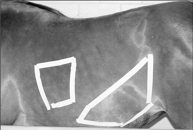
Picture of the left flank of a horse, displaying the regions where the stomach (cranial) and the large colon (caudal) were visualized in this study.
Figure 2.
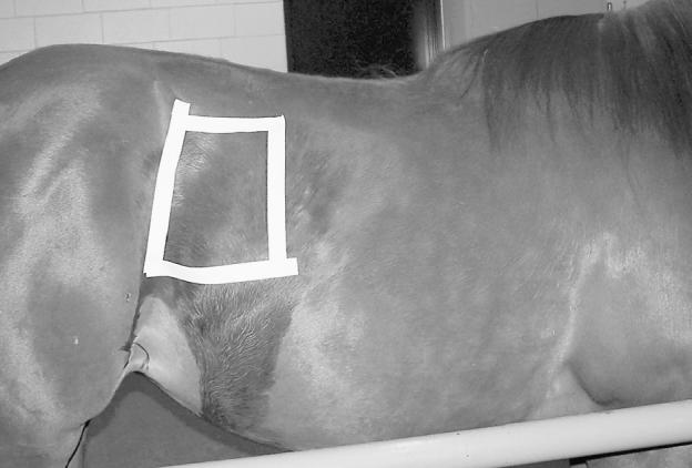
Picture of the right flank of a horse, displaying the region where the base of the cecum was visualized in this study.
The large intestine was examined ventrally on the left side of the abdomen and the ventral colon was identified by the presence of sacculations, its anatomical location, and its ultrasonographic appearance (bright hyperechoic line and the inability to identify the entire circumference of its wall) (Figure 3). The cecal body was identified by its having a similar appearance to the ventral large intestine and by its position in the right caudal dorsal quadrant, adjacent to the body wall (Figure 4). The jejunum was identified as loops of intestine of considerably smaller diameter than the large intestine. With jejunum, the entire circumference of hypoechoic wall could be seen (Figure 5). The stomach was scanned from the 9th to the 12th intercostal space on the left side and identified by the thickness of its wall and its smooth, bright, round appearance (Figure 6). Subjective activity of the jejunum, cecum, and large intestine was classified as either active or inactive.
Figure 3.
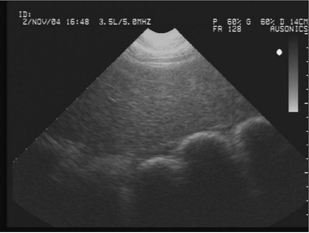
Ultrasonograph of the left ventral colon deep to the spleen obtained with the 3.5 MHz probe. The bright hyperechoic line indicates the luminal contents causing acoustic shadowing.
Figure 4.
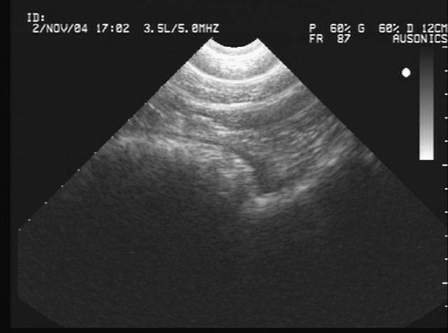
Ultrasonograph of the base of the cecum adjacent to the body wall showing sacculations in the cecal wall and its hyperechoic luminal contents.
Figure 5.
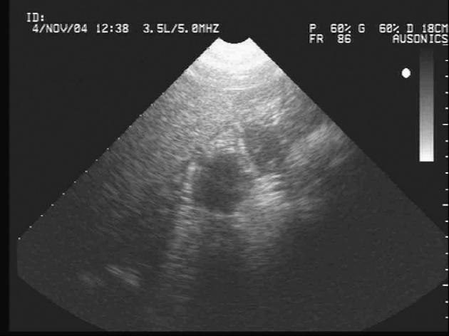
Ultrasonograph of 2 loops of jejunum identified by their small diameter and the ability to visualize the wall’s entire circumference.
Figure 6.
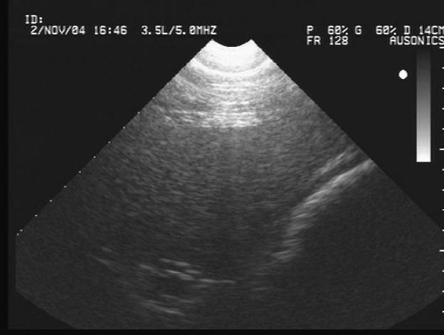
Ultrasonograph of the stomach adjacent to the liver. The stomach is identified by the thickness of its wall, which is much thicker than that of the adjacent large colon.
Pulsed-wave Doppler ultrasound was used when possible to assess progressive activity of the jejunum, by placing the sample volume within the lumen of the bowel. The sample volume is the area being examined for the Doppler shift and it is identified on the ultrasonograph as the area between the 2 parallel lines on the B mode display (Figure 7). The resultant tracing associated with the Doppler technique indicates the velocity of flow of the fluid in the area within the sample volume. The Doppler angle was between 30° and 60° (angle correction software built into the program and used in all assessments), and the sample volume size was 5 mm. Doppler recordings were taken only when the same jejunal segment was visible for sufficient duration to accurately set the sample volume to obtain both B mode and Doppler readings. In 1 sedated horse in trial 1, Doppler examination was never possible due to the extreme movement of the loop of bowel and the inability to accurately place the sample volume. No data were collected for stomach, cecum, or large intestine, as the gas in these organs prevents readings from being obtained. Doppler patterns were classified as either peristaltic (Figure 7) or nonperistaltic (Figure 8). These classifications were based on the human literature (5) and on previous results obtained by the investigators using direct jejunal contact in experimental horses undergoing nonsurvival exploratory laparotomy. When crescendo/decrescendo wave forms or excursions from baseline were observed (indicating fluid flow towards or away from the transducer), activity was classed as peristaltic. When no deviation from the baseline was detected, the intestine was classified as having no peristaltic activity. In preliminary studies of active intestine, local activity (segmentation type pattern) did not result in a Doppler tracing. Patterns of single spikes on the Doppler tracing or other movement artifacts (waves lacking crescendo/decrescendo pattern) and reverberations were excluded from analysis. Three recordings were taken at each site of visible jejunum. If mixed patterns were detected on these different recordings (one recording active and another inactive), the intestine was classed as being active.
Figure 7.
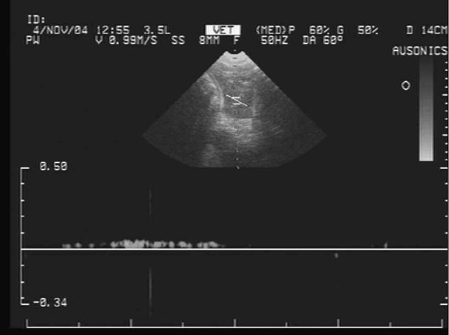
Ultrasonograph of pulsed-wave Doppler activity of jejunum. The sample volume is the set of parallel lines in the B mode image centered over the lumen of a loop of jejunum and the Doppler display shows peristaltic activity as a deviation of the tracing from the baseline. The horizontal axis represents 4 s.
Figure 8.
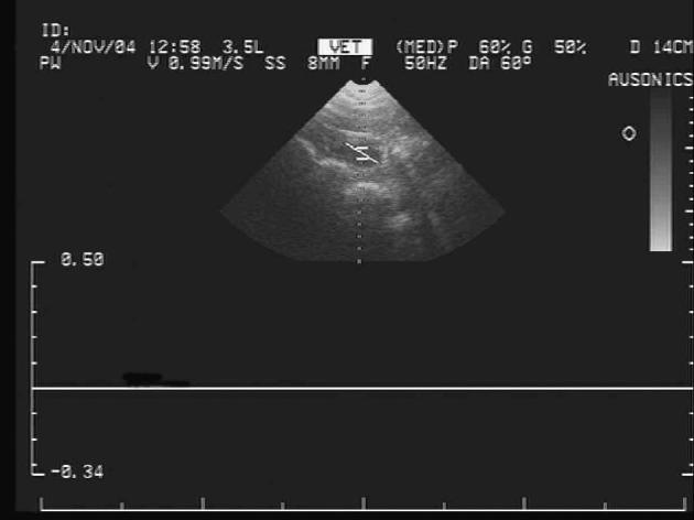
Ultrasonograph of pulsed wave Doppler activity of jejunum. The Doppler display shows no peristaltic activity. The horizontal axis represents 4 s.
Statistics
Comparisons were made between the pairs of each trial, between the on and off feed groups, between the 2 off feed groups, and between Doppler and B mode activity descriptions. Nominal and ordinal data (intestinal contents, B mode activity, Doppler activity) were analyzed by using Chi-squared tests. Numerical data (contraction rate) were analyzed by using paired t-tests, after normal distribution was verified by plotting data against a normal curve. B mode and Doppler activity patterns were evaluated for association independently by 2 of the authors (CM, EM) by using McNemar’s test, and a kappa coefficient was calculated for measurement of agreement. Significance was set at P < 0.05.
Results
Trial 1 (fed horses with and without sedation with xylazine) was completed with ultrasonographic data collected for 10 horses. Trial 2 (fasted baseline control and following nasogastric intubation) was completed using only 9 horses, due to the inability to intubate the 10th horse. Horses were fasted for an average of 15.8 ± 4.9 h (range 8 to 24 h). Trial 3 (fasted horses with and without sedation) was completed using 10 horses, with an average fasting time of 19.3 ± 4.5 h (range 13 to 24 h, all horses except 1 were a minimum of 14 h).
Stomach
In healthy fed horses, the stomach was seen between the 9th and 12th intercostal spaces on the left side, proximal to the costochondral junction. No contractions were observed and the stomach always contained gas. When these same horses were fasted, the stomach was not identified in its usual position (8), instead it was identified ventral to the costochondral junction. Following placement of an indwelling nasogastric tube, the stomach was again visible in its normal location dorsal to the costochondral junction. Two horses spontaneously refluxed after the tube had been in place for 30 min. Xylazine sedation did not cause any observable alterations in the position or activity of the stomach in either fed or fasted horses.
Jejunum (Table 1)
Table 1.
Ultrasonographic measurements of the jejunum in 10 healthy horses in trial 1 (fed versus fed and sedated), trial 2 (fasting), and trial 3 (fasted versus fasted and sedated), except for trial 2 (fasting and nasogastric intubation [NGT]) where 9 horses were used
| B mode activity | Doppler activity | |||||
|---|---|---|---|---|---|---|
| Group | Visibility | Contractions/minute (χ̄) | n | Grade | n | Grade |
| Fed (Trial 1) | 10% | NM | 1 | Active | 1 | ND |
| Fed and sedated (Trial 1) | 10% | 0.00 (n = 1) | 1 | Inactive | 1 | NM |
| Fasted (Trial 2) | 60% | 12.27, sχ̄ = 6.65 (n = 4) | 0 | Inactive | 1 | ND |
| 6 | Active | 4 | Inactive | |||
| Fasted and NGT (Trial 2) | 67% | 24.54, sχ̄ = 13.31 (n = 4) | 1 | Inactive | 1 | Active |
| 5 | Active | 5 | Inactive | |||
| Fasted (Trial 3) | 90% | 3.15, sχ̄ = 1.09 (n = 5) | 0 | Inactive | 0 | Active |
| 8 | Active | 9 | Inactive | |||
| Fasted and sedated (Trial 3) | 100% | 6.30, sχ̄ = 2.18 (n = 5) | 5 | Inactive | 4 | Active |
| 5 | Active | 6 | Inactive | |||
Nonpercentile values expressed as mean (χ̄) and their standard error (sχ̄)
NM — not measured; ND — nondiagnostic
In healthy fed horses, the jejunum was observed in only 1/10 (10%) horses. In this horse, the jejunum was active and observed in the left ventral abdomen, but Doppler assessment was not possible due to the inability to accurately place and maintain the sample volume. When these horses were fasted, the jejunum was visualized more frequently (60% to 90% versus 10%; P < 0.05 for trial 2 and P < 0.005 for trial 3) and was usually located ventrally in the abdomen. Jejunal visibility increased when fed and sedated horses were compared with fasted and sedated horses (P < 0.0001). Nasogastric intubation in fasted horses and xylazine sedation in fed horses did not cause any visible alterations. When fasted horses were sedated, the B mode and Doppler assessments of jejunal activity were decreased (P < 0.05). Doppler evaluation and determination of contraction rate in studies of the jejunum were frequently difficult, due to intestinal position or movement of intestine or patient during the examination.
Through the course of the study, Doppler and B mode results were both obtained on the same jejunal segment for a total of 29 examinations. These results matched in their assessment of jejunal activity in 24 examinations (83%). Association between B mode and Doppler assessments displayed moderate agreement (kappa coefficient = 0.51).
Cecum (Table 2)
Table 2.
Ultrasonographic measurements of the cecum in 10 healthy horses in trial 1 (fed versus fed and sedated), trial 2 (fasting), and trial 3 (fasted versus fasted and sedated), except for trial 2 (fasting and nasogastric intubation [NGT]) where 9 horses were used
| B mode activity | ||||
|---|---|---|---|---|
| Group | Visibility | Contractions/minute (χ̄) | n | Grade |
| Fed (Trial 1) | 100% | 3.97, sχ̄ = 0.58 | 1 | Inactive |
| 9 | Active | |||
| Fed and sedated (Trial 1) | 100% | 2.66, sχ̄ = 0.84 | 1 | Inactive |
| 9 | Active | |||
| Fasted (Trial 2) | 100% | 1.80, sχ̄ = 0.45 | 2 | Inactive |
| 8 | Active | |||
| Fasted and NGT (Trial 2) | 100% | 3.18, sχ̄ = 0.74 | 2 | Inactive |
| 7 | Active | |||
| Fasted (Trial 3) | 100% | 2.43, sχ̄ = 0.48 | 0 | Inactive |
| 10 | Active | |||
| Fasted and sedated (Trial 3) | 100% | 0.60, sχ̄ = 0.19 | 6 | Inactive |
| 4 | Active | |||
Nonpercentile values expressed as mean (χ̄) and their standard error (sχ̄)
In fed horses, the cecum was always visible and activity was always observed. In fasted horses (trial 2 and 3), cecal contraction rate was decreased (P < 0.05) when compared with that in the fed group. Gas was present in the cecum of the horses during all trials, with the exception of trial 3, when the contents changed and fluid was identified within the cecum in 5/10 (50%) horses. Nasogastric intubation in fasted horses and xylazine sedation in fed horses did not cause any visible alterations in the cecum. Sedation in fasted horses significantly decreased the cecal contraction rate (P = 0.007) and B mode activity, as compared with those of horses that had only been fasted (P < 0.005).
Large intestine (Table 3)
Table 3.
Ultrasonographic measurements of the large intestine in 10 healthy horses in trial 1 (fed versus fed and sedated), trial 2 (fasting), and trial 3 (fasted versus fasted and sedated), except for trial 2 (fasting and nasogastric intubation [NGT]) where 9 horses were used
| B mode activity | ||||
|---|---|---|---|---|
| Group | Visibility | Contractions/minute (χ̄) | n | Grade |
| Fed (Trial 1) | 100% | 3.05, sχ̄ = 1.00 | 2 | Inactive |
| 8 | Active | |||
| Fed and sedated (Trial 1) | 100% | 2.33, sχ̄ = 0.74 | 3 | Inactive |
| 7 | Active | |||
| Fasted (Trial 2) | 100% | 1.65, sχ̄ = 0.39 | 3 | Inactive |
| 7 | Active | |||
| Fasted and NGT (Trial 2) | 100% | 1.93, sχ̄ = 0.74 | 2 | Inactive |
| 7 | Active | |||
| Fasted (Trial 3) | 100% | 0.63, sχ̄ = 0.27 | 4 | Inactive |
| 6 | Active | |||
| Fasted and sedated (Trial 3) | 100% | 0.53, sχ̄ = 0.22 | 6 | Inactive |
| 4 | Active | |||
Nonpercentile values expressed as mean (χ̄) and their standard error (sχ̄) The only significant differences seen between fasted examinations in trial 2 and trial 3 were in large intestinal contractions per minute
In fed horses, the large intestine was always visible and activity was always observed. The contraction rate decreased following fasting, but this was only significant for trial 3 (P = 0.03). In trial 3, the large intestinal contraction rate was further decreased (0.63, sχ̄= 0.27 versus 1.65, sχ̄= 0.39 per min; P = 0.02) when compared with that in trial 2. Placement of a nasogastric tube or xylazine sedation did not cause any observable alterations. The large intestine always contained gas.
Discussion
In healthy horses, fasting caused significant, sonographically observable alterations in intestinal activity and visibility throughout the length of the gastrointestinal tract examined. The effects of xylazine were dependent on feeding status, with minimal changes being observed in fed horses but with significant effects observed in fasted animals. An indwelling nasogastric tube produced minimal alterations in jejunal, cecal, or colonic visibility and activity.
Stomach activity throughout this experiment appeared to be non existent, regardless of group, most likely because it is the greater curvature of the fundic and proximal body that can be visualized. This region of the stomach is involved mainly with receptive relaxation rather than active contractions (9). In fasted horses, decreased ingesta volume may be responsible for the unexpected change in position of the stomach. Placement of a nasogastric tube is a vital component of treatment and evaluation of colic. Very little information is available regarding the consequences of this with regards to effects on the remainder of the intestinal tract. In fasted horses, it allowed the stomach to be visualized in its normal position, where it had not been seen prior to intubation. This may have been due to local irritation and subsequent alterations in filling caused by the indwelling nasogastric tube (10,11).
Jejunal sonographic activity in fasted animals differed significantly from that in fed animals. Fasted horses had increased jejunal visibility and decreased activity. The increase in visibility may have occurred due to emptying of the colon and cecum and should be considered a normal finding in fasted horses. When fasted horses were sedated with xylazine, further decreases in jejunal activity were observed. This is consistent with the generally expected effects of xylazine described in the literature (12,13). However, xylazine sedation in fed horses was found to have little effect on jejunal activity, in agreement with results obtained from fed horses published elsewhere (14). The difference may reflect the use of fasted horses in most motility studies (12,13,15). Xylazine may still be able to decrease electrical and muscular activity, but the presence of food may stimulate the local, gastrocolic, and enterocolic reflexes sufficiently to prevent any inhibition from being observed ultrasonographically.
In fed horses, the cecum and large intestine were routinely visualized. Contraction rates of the cecum and large intestine were consistent with those obtained in other ultrasonographic studies (4,7) and in pressure wave studies (16) but tended to be much less than those obtained in electrode studies (17). It may be that certain electrical patterns are not reflected in observable activity or that lower level activity patterns were missed. Fasted horses had decreased activity of the cecum and large intestine. Decreases in large intestinal and cecal activity may have been due to depression of the gastrocolic and enterocolic reflexes or to changes in content type that decreased stimulation of local receptors. Ingesta were still present within these organs, so a lack of local peristaltic reflexes seems unlikely. With fasting, cecal contents in a number of horses also changed from gas to fluid, possibly reflecting less substrate available for fermentation and therefore less gas production. The cecum seemed more sensitive to fasting than did the large intestine. This may be related to more rapid transit times, less complex mixing activity, or both, in the cecum than in the colon (17). Colonic activity was not significantly affected by fasting or sedation. However, cecal activity was decreased further with fasting and sedation than with fasting alone. Similar to what was observed in the jejunum, effects of sedation were minimal in fed horses.
Continuous Duplex Doppler ultrasound is used in human medicine to evaluate intestinal peristalsis and cardiac blood flow. The use of Doppler ultrasound was included as a potential identifier of equine intestinal peristalsis. In only one other study has differentiation between peristaltic and mixing contractions in equine jejunum with the use of ultrasonography been reported (4). In that study, transrectal ultrasonography allowed longitudinal sections of intestine to be visualized consistently, enabling propulsive motility to be identified by peristaltic contractions and movement of ingesta aborally. Jejunal Doppler readings were obtained in 30/33 (91%) examinations when jejunum was identified. This technique is limited in its clinical use to whenever jejunum is visualized, but it should provide data in the majority of horses that have been fasted or in some cases of colic. Measurement errors were hard to eliminate in equine patients when transcutaneous ultrasonography was used, as patient movement and the depth and movement of the tissues being examined resulted in problems in keeping the Doppler focused on the bowel.
In this study, B mode and Doppler patterns were highly correlated, with results matching in 83% of the examinations. We made no attempt to quantify Doppler tracings but examined patterns only. Further studies may enable more information to be obtained, especially regarding whether Doppler is more accurate than B mode ultrasound at differentiating between mixing and peristaltic activity. Increasing operator experience with Doppler ultrasonography, using higher powered probes, and technological advances in equipment used for equine patients should lead to greater accuracy and sensitivity. The need for Duplex Doppler (simultaneous real time B mode and Doppler ultrasound) was highlighted in this study by the difficulty in obtaining accurate Doppler readings in some animals.
In summary, ultrasonography is useful for evaluating gastrointestinal activity. The correlation of our results to previous reports in the literature on the effects of xylazine in fasted horses and to pressure wave measurements suggest that ultrasonography is a viable tool for noninvasive quantification of intestinal activity. Feeding status and sedation must be taken into consideration in horses undergoing sonographic gastrointestinal evaluation, as fasting related changes in stomach and jejunal location can be misleading. However, in fed horses, xylazine sedation of 0.3 to 0.4 mg/kg BW, IV, should not significantly alter activity. Nasogastric intubation did not appear to affect sonographic findings other than stomach visualization. Finally, quantification of intestinal activity and investigation of Doppler ultrasonography have the potential to further enhance the usefulness of ultrasound as a diagnostic tool in the horse. CVJ
Footnotes
Reprints will not be available from the authors.
This project was supported by the University of Minnesota Equine Center with funds provided by the Minnesota Racing Commission, Minnesota Agricultural Experimental Station and contributions by private donors.
References
- 1.McGladdery AJ. Ultrasonography as an aid to the diagnosis of equine colic. Equine Vet Educ. 1992;4:248–251. [Google Scholar]
- 2.Klohnen A, Vachon AM, Fischer AT. Use of diagnostic ultrasonography in horses with signs of acute abdominal pain. J Am Vet Med Assoc. 1996;209:1597–1601. [PubMed] [Google Scholar]
- 3.Braun U, Marmier O. Ultrasonographic examination of the small intestine of cows. Vet Rec. 1995;136:239–244. doi: 10.1136/vr.136.10.239. [DOI] [PubMed] [Google Scholar]
- 4.Freeman SL, England GCW. Effect of romifidine on gastrointestinal motility, assessed by transrectal ultrasonography. Equine Vet J. 2001;33:570–576. doi: 10.2746/042516401776563436. [DOI] [PubMed] [Google Scholar]
- 5.Gimondo P, La Bella A, Mirk P, Torsoli A. Duplex-Doppler evaluation of intestinal peristalsis in patients with bowel obstruction. Abdom Imaging. 1995;20:33–36. doi: 10.1007/BF00199641. [DOI] [PubMed] [Google Scholar]
- 6.Gimondo P, Mirk P. A new method for evaluating jejunal motility using duplex Doppler sonography. Am J Roentgenol. 1997;168:187–192. doi: 10.2214/ajr.168.1.8976944. [DOI] [PubMed] [Google Scholar]
- 7.Reef VB. Equine Diagnostic Ultrasound. 1st ed. Philadelphia: WB Saunders, 1998:273–363.
- 8.Cannon JH, Andrews A. Ultrasound of the equine stomach. Proc Am Assoc Equine Pract. 1995;41:38–39. [Google Scholar]
- 9.Ehrlein HJ, Akkermans LMA. Gastric emptying. In: Akkermans LMA, Johnson AG, Read NW, eds. Gastric and Gastroduodenal Motility. 1st ed. London: Praeger Publ, 1984:74–84.
- 10.Dabareiner RM. Treatment after abdominal surgery. In: White NA, Moore JN, eds. Current Techniques in Equine Surgery and Lameness. 2nd ed. Philadelphia: WB Saunders, 1998:295–297.
- 11.Mair TS. Small intestinal obstruction caused by a mass of feedblock containing molasses in 4 horses. Equine Vet J. 2002;34:532–536. doi: 10.2746/042516402776117719. [DOI] [PubMed] [Google Scholar]
- 12.Lester GD, Merritt AM, Neuwirth L, Vetro-Widenhouse T, Steible C, Rice B. Effect of α2 -adrenergic, cholinergic and nonsteroidal anti-inflammatory drugs on myoelectric activity of ileum, cecum, and right ventral colon and on cecal emptying of radiolabeled markers in clinically normal ponies. Am J Vet Res. 1998;59:320–327. [PubMed] [Google Scholar]
- 13.Merritt AM, Burrow JA, Hartless CS. Effect of xylazine, detomidine, and a combination of xylazine and butorphanol on equine duodenal motility. Am J Vet Res. 1998;59:619–623. [PubMed] [Google Scholar]
- 14.Merritt AM, Campbell-Thompson ML, Lowrey S. Effect of xylazine treatment on equine proximal gastrointestinal tract myoelectrical activity. Am J Vet Res. 1989;50:945–949. [PubMed] [Google Scholar]
- 15.Adams SB, Lamar CH, Masty J. Motility of the distal portion of the jejunum and pelvic flexure in ponies: Effects of six drugs. Am J Vet Res. 1984;45:795–799. [PubMed] [Google Scholar]
- 16.Lowe JE, Sellers AF, Brondum J. Equine pelvic flexure impaction. A model used to evaluate motor events and compare drug response. Cornell Vet. 1980;70:401–412. [PubMed] [Google Scholar]
- 17.Merritt AM, Panzer RB, Lester GD, Burrow JA. Equine pelvic flexure myoelecric activity during fed and fasted sttes. Am J Physiol. 995;269:262–268. doi: 10.1152/ajpgi.1995.269.2.G262. [DOI] [PubMed] [Google Scholar]


