
FIGURE 1.
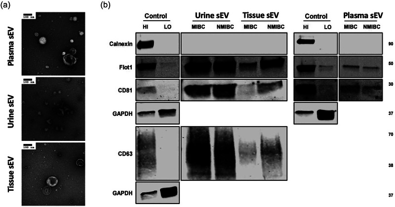
(a) TEM images confirming the presence of negative‐stained sEVs separated from plasma, urine and tissue explants, seen as cup‐shaped vesicles. Scale bars are 100 nm. (b) Western blots of flotillin‐1, CD63, CD81, and calnexin for sEVs separated from plasma, urine and tissue explants. MCF7 membrane and cytosolic protein fractions served as positive and negative controls for flotillin‐1, CD63, CD81 and calnexin.