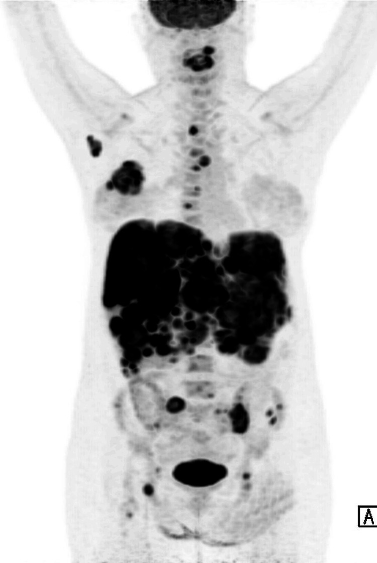
Figure 2. Positron emission tomography examination (PET) .
A hypermetabolic right breast mass is noted at 3 cm from the nipple. Multifocal liver metastases were noted diffusely infiltrating both hepatic lobes. Multiple focal bony metastases were noted in the axial and appendicular skeleton as described. Of note is the involvement of the cervical spine with vertebral plana deformity at C3 with bony retropulsion and compression of the spinal cord at C3/4 level.

