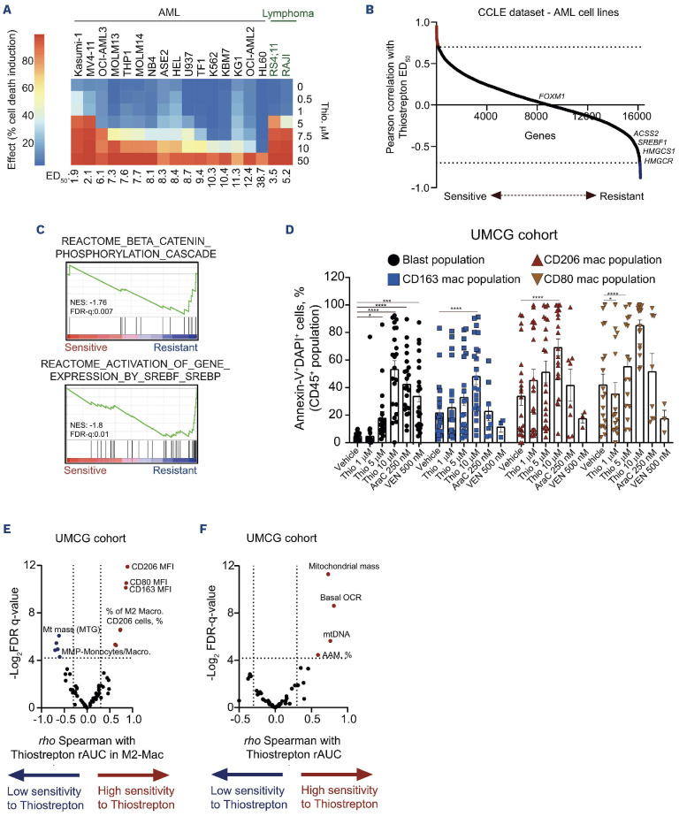
Figure 2.
Thiostrepton can target both the leukemic cells and the tumor microenvironment in acute myeloid leukemia. (A) Median effective dose (ED50) of thiostrepton in a panel of acute myeloid leukemia (AML) and B-cell acute lymphoblastic leukemia (B-ALL)/lymphoma cell lines. (B) Hockey stick plot displaying the correlation between RNA-sequencing data from AML cell lines (CCLE dataset) and thiostrepton sensitivity. Expression of SREBP target genes was correlated with thiostrepton insensitivity (C) Gene set enrichment analysis (GSEA) using the Pearson correlations depicted in (B) normalized enrichment score (NES) and false discover rate (FDR-q) are indicated. (D) Ex vivo drug-induced apoptosis of thiostrepton in a set of 22 primary AML samples (left panel). Cells were treated for 72 hours, and apoptosis was evaluated by Annexin-V/ DAPI staining by flow cytometry. Leukemic blast cells were analyzed in the CD45dimCD34+CD117+ population, while macrophages were analyzed in the CD45highCD14+CD163+ population. (E, F) Volcano plot demonstrating the Spearman correlation between the ex vivo thiostrepton-induced apoptosis (reported as reverse area under the curve [rAUC]) on M2 macrophage (E) and AML blasts (F) treated ex vivo in our set of primary AML samples (N=22 samples). Blue dots indicate significant negative correlation and red dot indicate a positive correlation. Bar graphs represent the mean ± standard error of the mean of at least 3 independent experiments. *P<0.05, **P<0.01, ***P<0.001. UMCG: University Medical Center Groningen.

