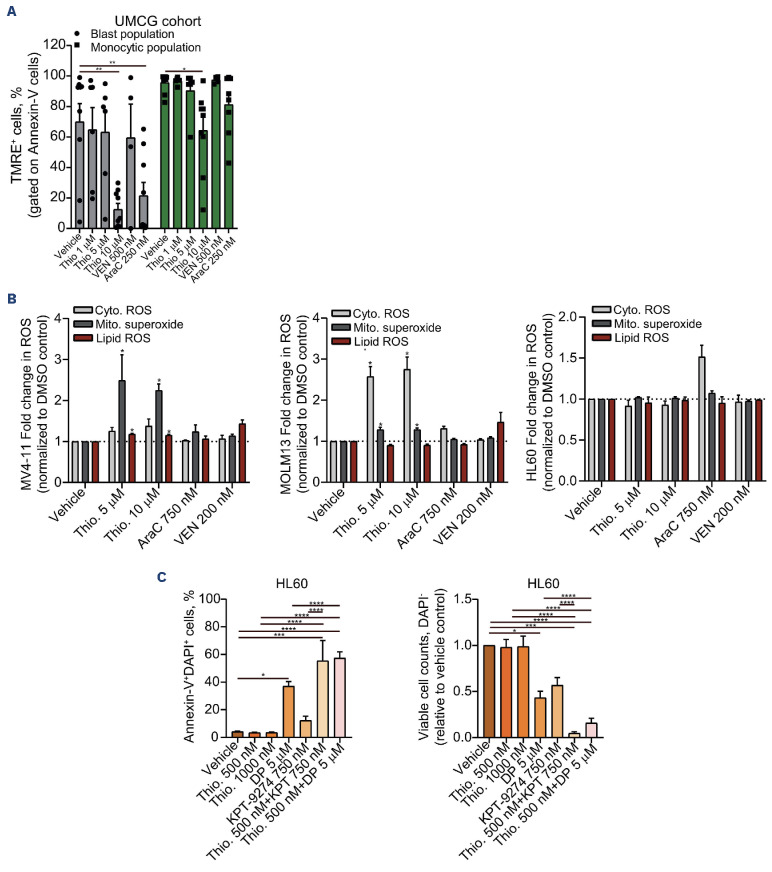
Figure 3.
Decreased mitochondrial membrane potential and increased reactive oxygen species upon thiostrepton treatment. (A) Mitochondrial membrane potential was detected by flow cytometry in acute myeloid leukemia (AML) blasts, gated on CD45+CD34+ (or CD117+ for NPM1 mutant AML)/Annexin V- and the myeloid-associated monocytic population (gated on SSChighCD45highCD163+ cells) using the TMRE staining method. Cells were treated with vehicle dimethyl sulfoxide [DMSO], cytarabine (AraC, 250 nM), venetoclax (VEN, 500 nM), or thiostrepton (1-5-10 mM) for 72 hours (hrs). Bar graphs represent the mean ± standard error of the mean, each point represents a patient sample. (B) Fold change in lipid reactive oxygen species (ROS) (C11-BODIPY), cytoplasmic ROS (DCFDA), and mitochondrial superoxide levels (MitoSOXTM) in MV4-11, MOLM13 and HL60 cells treated with thiostrepton (5-10 mM), cytarabine (AraC, 750 nM) or venetoclax (VEN, 200 nM) for 72 hrs; N=4 independent experiments. (C) Apoptosis levels (left panel) and viable cell counts (relative to vehicle control, right panel) measured by flow cytometry in HL60 cells treated with vehicle or with increasing concentrations of thiostrepton (0.5-1 mM) and/or dipyridamole (DP, 5 mM) or KPT-9274 (750 nM) for 72 hrs using an APC-Annexin V/DAPI staining method. Bar graphs represent the mean ± standard error of the mean of at least 3 independent experiments. *P<0.05, **P<0.01, ***P<0.001. UMCG: University Medical Center Groningen.

