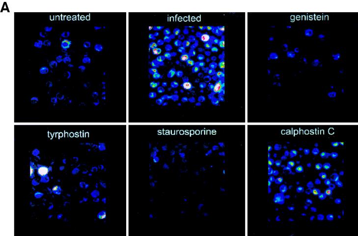
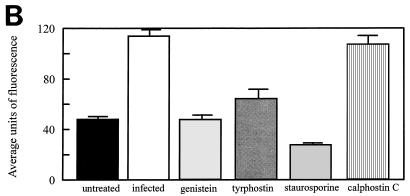
FIG. 5.
Immunofluorescent staining of phosphotyrosine proteins present during L. pneumophila entry into human monocytes. (A) Observation was done by confocal microscopy. (B) Quantitation was done with Meridian software. Infected cells were exposed to L. pneumophila for 15 min. Cells were pretreated with genistein 15 min prior to infection or with tyrphostin, staurosporine, or calphostin C 60 min prior to infection. Cells were stained with antiphosphotyrosine antibody 4G10 and with a tetramethyl rhodamine isocyanate-conjugated goat anti-mouse antibody. Results are reported as means ± the standard errors of the means for three experiments.


