
Figure 1.
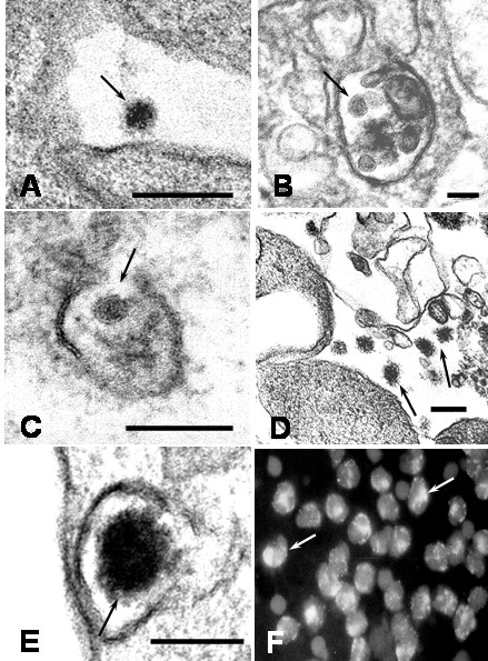
Electron microscope morphological observations of DEN2 virus particles. A) Typical viral particle in the extracellular environment (arrow; bar: 200 nm). B) Viral particles engulfed in an intracytoplasmic vacuole (arrow; bar: 50 nm). C) Membrane disruption of a vesicle containing a virus (arrow; bar: 100 nm). D) Fuzzy coated viral particles occur in the extracellular space (arrows; bar: 200 nm) E) A fuzzy coated viral particle showing an envelope with projections (arrow; bar: 100 nm). F) Immunofluorescence staining of DEN2 viral antigens at 4 h of culture. A diffuse and patchy pattern of fluorescence was observed in the cytoplasm (arrows). × 1000.