
Figure 10.
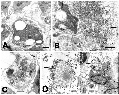
Ultrastructural features of apoptotic cells in DEN2 virus-infected monocytes at 6 hours. A) Swelling of organelles and membrane compartments in an apoptotic cell (bar: 1 μm). B) Release of cellular content from a swelling apoptotic cell. Note numerous viral particles (arrow) probably already present in the extracellular space (bar: 1 μm). C) Advance phase of cellular swelling (ghost cell) showing disruption of plasma membrane (arrow; bar: 1 μm). D) Ghost cell surrounding by numerous viral particles (arrow; bar: 500 nm). E) Monocyte engulfing cellular debris (arrow). Note the presence of a phagosome containing partial digested cellular material (bar: 1 μm).