
Figure 4.
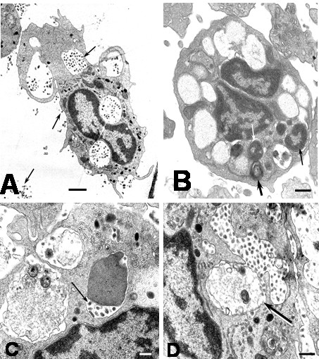
Ultrastructural features of DEN2 virus-infected monocytes. A) DEN2 virus-infected monocytes after 2 hours of infection. Observe the presence of virus particles in the extracellular space, on cellular plasma membrane and inside cytoplasmic vacuoles (arrows; bar: 1 μm). B) Monocyte showing huge empty vacuoles and vacuoles containing nuclear debris and myelin structures at 4 hours of culture (arrows; bar: 500 nm). C) Monocyte showing cytoplasmic phagosomes containing cellular debris and viral particles (arrow; bar: 200 nm). D) A huge vacuole containing numerous viral particles and cellular debris (arrow; bar: 500 nm).