
Figure 7.
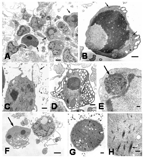
Ultrastructural features of apoptotic cells in DEN2 virus-infected monocytes at 4 hours. A) The typical features of apoptosis are observed in several monocytes (arrows; bar: 2 μm). B) Apoptotic cell showing cellular shrinkage, nuclear condensation and bundles of microfibrils (arrows; bar: 500 nm). C) Monocyte with dense remnant nucleus and surface blebbing (arrow; bar 500 nm). D) Apoptotic cell showing intense cytoplasmic vacuolization (bar: 500 nm). E) Phagosome in the cytoplasm of apoptotic cell (arrow; bar: 200 nm). F) Nuclear fragmentation in apoptotic cell (arrow). Note beside a healthy monocyte (bar: 2 μm). G) Segment of apoptotic cell showing numerous bundles of cytoplasmic fibrils (bar: 200 nm). H) Bundles of microfibrils (arrow) in the cytoplasm of apoptotic cell (bar: 100 nm).