
Figure 9.
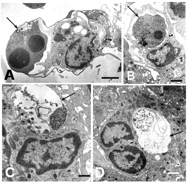
Ultrastructural features of apoptotic cells in DEN2 virus-infected monocytes. Different phases of phagocytosis and digestion of apoptotic cells. A) Engulfment of apoptotic cell (arrow) by a monocyte (bar: 2 μm). B) A huge phagosome containing a morphological intact apoptotic cell (arrow; bar: 1 μm). C and D show phagosomes (arrows) containing a partial digested apoptotic cells (C bar: 500 nm; D bar: 1 μm).