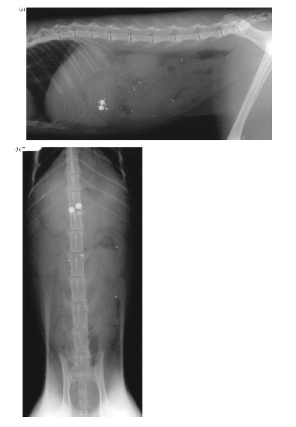
Fig 1.

Lateral (A) and ventrodorsal (B) abdominal radiographs of a cat with perforated gastric ulcers of undetermined cause (case 3). Note the loss of serosal detail indicative of peritoneal fluid accumulation secondary to generalised peritonitis. Retention of the barium-impregnated polyethylene spheres in the stomach 36 h after administration was suggestive of delayed gastric emptying or a pyloric outflow obstruction.
