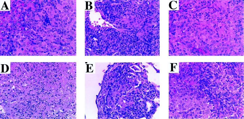
FIG. 3.
Higher-magnification photomicrographs of some of the lesions depicted in Fig. 2, demonstrating cytological details. (A) PBS control. Scattered lymphocytes are admixed with epithelioid macrophages. (B) BCG. Numerous lymphocytes are present throughout the section. There is no caseation. (C) MPL control. Note the relative paucity of lymphocytes amid numerous foamy macrophages. (D) MPL-CFP. This section demonstrates an area of central caseation, the same lesion seen in the lungs of MPL–CFP–IL-12-vaccinated animals (higher magnification not included). (E) MPL–CFP–IL-2. Note the similarity to the lesion in the BCG control (B). This is essentially the same lesion observed in MPL–CFP–IL-2–IL-12-vaccinated animals (higher magnification not included). (F) DNA-Ag85A. Relatively numerous lymphocytes are admixed with fields of epithelioid macrophages. Hematoxylin and eosin stain; magnifications, ×156.

