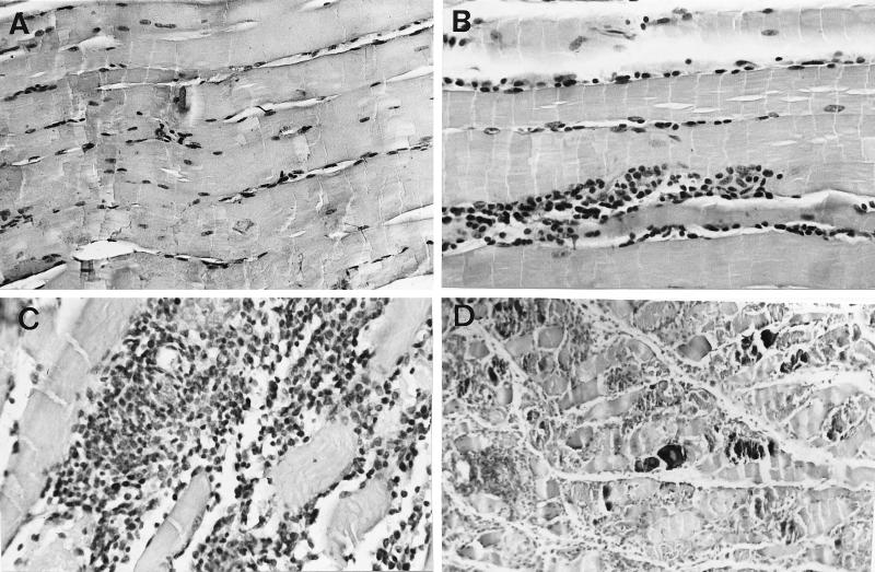
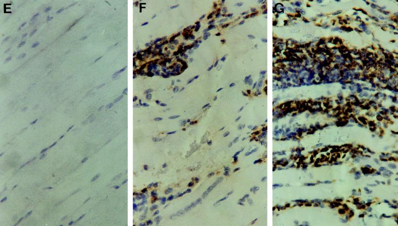
FIG. 5.
Hematoxylin-eosin stainings (A to D) and immunohistochemical study of CD45 expression (E to G) of skeletal muscles from TNFRp55−/− (A, C, D, and G) and WT (B, E, and F) mice at 0 (A and E) or 30 (B, C, D, F, and G) days after infection with T. cruzi (strain CA-I). Negative controls for immunohistochemical stainings done by incubation with normal rat serum on tissues from T. cruzi-infected mice showed no peroxidase staining (data not shown). It is important to note the dramatically increased density of inflammatory infiltrates (C and G) and the presence of necrosis and calcification (D) in tissues obtained from infected TNFRp55−/− mice.


