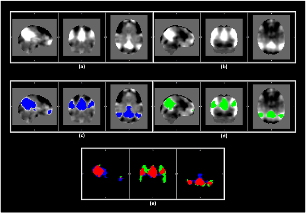
Fig. 2.
An example for the process of obtaining the same set of spatial maps to be used as the reference for GIG-ICA in both modalities. (a) and (b) show the default mode network (DMN) for rsfMRI and PASL data. (c) and (d) demonstrate the areas in blue and green associated with the DMN in rsfMRI and PASL data, respectively. (e) demonstrates these areas from (c) and (d) with the overlap indicated in red. (For interpretation of the references to colour in this figure legend, the reader is referred to the Web version of this article.)

