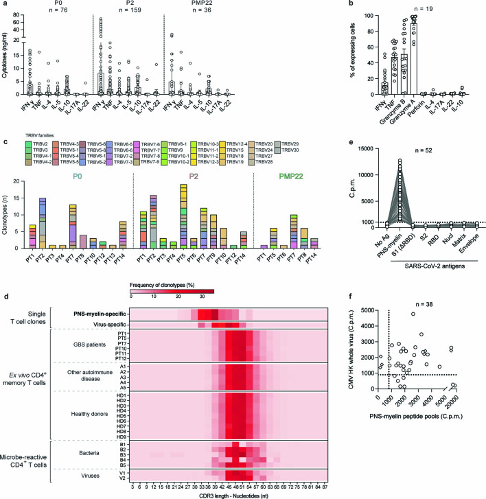
Extended Data Fig. 4. Characterization of autoreactive T cell clones in patients with GBS.
a,b, Cytokine production by P0-specific (n = 76 biologically independent samples), P2-specific (n = 159 biologically independent samples) and PMP22-specific (n = 36 biologically independent samples) CD4+ T cell clones derived from patients with GBS analysed in the 48-h-culture supernatants after stimulation or not with the self-antigen by bead-based multiplex assay (a) or intracellularly by intracellular FACS staining (b). Each dot represents an individual clone and bar height indicates mean value with s.d. (a) or s.e.m. (b). c, TCR Vβ gene usage of P0-, P2- and PMP22-specific TCRβ clonotypes isolated from the indicated patients with GBS. The y axis indicates the number of autoreactive TCRβ clonotypes. d, Heat map showing the percentage of clonotypes bearing the same CDR3β length defined by the number of nucleotides. The CDR3β lengths of TCRβ clonotypes from PNS-myelin-specific CD4+ T cells isolated from patients with GBS are shown in the top row (n = 166 from 13 PT) and were compared to the CDR3β lengths of (i) SARS-CoV-2-specific CD4+ T cell clones (virus-specific TCRβ clonotypes, n = 92 from 6 PT), (ii) ex vivo memory CD4+ T cells from the indicated patients with GBS, or patients with other autoimmune disorders (A) or healthy donors (C) and (iv) bacteria- (B) and viruses- (V) reactive CD4+ T cell CFSElow fractions enriched from in vitro stimulation assay. e,f, PNS-myelin specific CD4+ T cell clones (circles) from two post-COVID-19 patients with GBS (n = 52; PT12 and PT14) or from CMV-associated patients with GBS (n = 32; PT2 and PT3) as well as CMV-specific CD4+ T cell clones from PT2 (rhombus symbols, n = 6) were stimulated in the presence of irradiated autologous B cells pulsed with the PNS-myelin antigen or the indicated SARS-COV-2 antigens (e) or heat-inactivated human CMV (f). Shown are the C.p.m. values indicating the proliferation of autoreactive T cell clones measured after a 16-h pulse with [3H]-thymidine.

