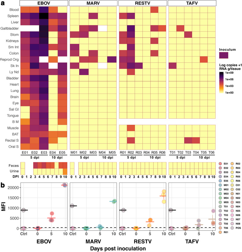
Fig. 2. Infection of Angolan free-tailed bats (AFBs) with filoviruses.
a Filovirus RNA copies/g of tissues or copies/ml of blood (rows) sampled from inoculated AFBs (columns) at day post-inoculation (dpi) 5 and 10. Bat identification numbers are noted below. Inoculum RNA load is shown as legend for reference. Blank spaces indicate unavailable samples (tissues, faeces and/or urine). Negative control bats are excluded Stom: Stomach; Sm Int: Small Intestine; Reprod Org: Testes or Uterus; Sk In: Inoculation point in Skin; Ly Nd: Lymph Node (cervical); Sal Gl: Salivary Gland; B M: Bone Marrow; BAT: Brown Adipose Tissue; S: Swab. b Serology (Luminex) of filovirus-inoculated (n = 22), positive (n = 2) and negative (n = 32) control AFBs. Circles show mean values ± SD (horizontal and error bars) of serum samples run in duplicate. Time point 0 shows pre-inoculation results, later re-analysed after euthanasia (5 and 10 dpi). EBOV and MARV positive controls from previously validated positive AFBs serum are shown as grey circles with black outline. Negative controls, including mock-inoculated (C01-C02) and bats kept in BSL4 caging (N01-N08) are shown in coloured circles with light-grey outline. The dashed line denotes the assay cut-off (mean value of negative controls + 3 SD). n = biologically independent animals. Source data are provided as a Source Data file.

