Fig. 5.
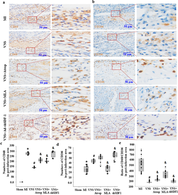
VNS altered M1/M2 macrophages in the peri-infarction area of myocardial infarction through m/n-AChR-SDF-1α. a, b Typical immunostaining image of CD68 (M1 macrophages) and CD206 (M2 macrophages) in the peri-infarction area of infarcted hearts treated with mancamylamine (MLA, 10 mg/kg, ip), atropine (Atrop, 10 mg/kg, ip) or SDF-1α knockdown by shRNA (Ad-shSDF-1) before VNS. Brownish yellow indicates CD68 or CD206; hematoxylin-stained nucleus. c, d Semiquantitative analysis of CD68 and CD206 numbers in the peri-infarct area of the infarcted hearts. *P < 0.05 vs. Sham; # P < 0.05 vs. MI; & P < 0.05 vs. VNS (n = 12). e Semiquantitative analysis of the ratio of CD68/CD206 levels in the peri-infarct area of the infarcted hearts. *P < 0.05 vs. Sham; # P < 0.05 vs. MI; & P < 0.05 vs. VNS (n = 12)

