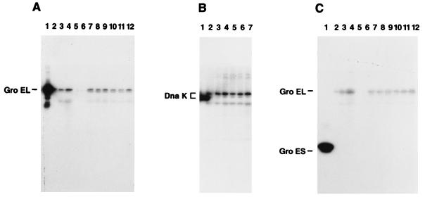
FIG. 1.
Detection of GroEL, DnaK, and GroES homologs. Western immunoblots of polyclonal rabbit antiserum against cell lysate proteins. (A) Lanes: 1, purified E. coli GroEL protein; 2, S. aureus maintained at 30°C; 3, S. aureus maintained at 37°C; 4, S. aureus shifted from 30 to 37°C; 5, cycloheximide-treated McCoy cells; 6, heat-killed bacteria added to McCoy cells; 7 to 9, S. aureus grown at 37°C and added to McCoy cells at 37°C (1, 2, and 3 h postinfection, respectively); 10 to 12, S. aureus grown at 30°C and added to McCoy cells at 37°C (1, 2, and 3 h postinfection, respectively). (B) Lanes: 1, purified E. coli DnaK protein; 2, S. aureus maintained at 30°C; 3, S. aureus maintained at 37°C; 4, S. aureus shifted from 30 to 37°C; 5 to 7, S. aureus grown at 37°C and added to McCoy cells at 37°C (1, 2, and 3 h postinfection, respectively). (C) Lanes: 1, purified E. coli GroES protein; 2, S. aureus maintained at 30°C; 3, S. aureus maintained at 37°C; 4, S. aureus shifted from 30 to 37°C; 5, cycloheximide-treated McCoy cells; 6, heat-killed bacteria added to McCoy cells; 7 to 9, S. aureus grown at 30°C and added to McCoy cells at 37°C (1, 2, and 3 h postinfection, respectively); 10 to 12, S. aureus grown at 37°C and added to McCoy cells at 37°C (1, 2, and 3 h postinfection, respectively).

