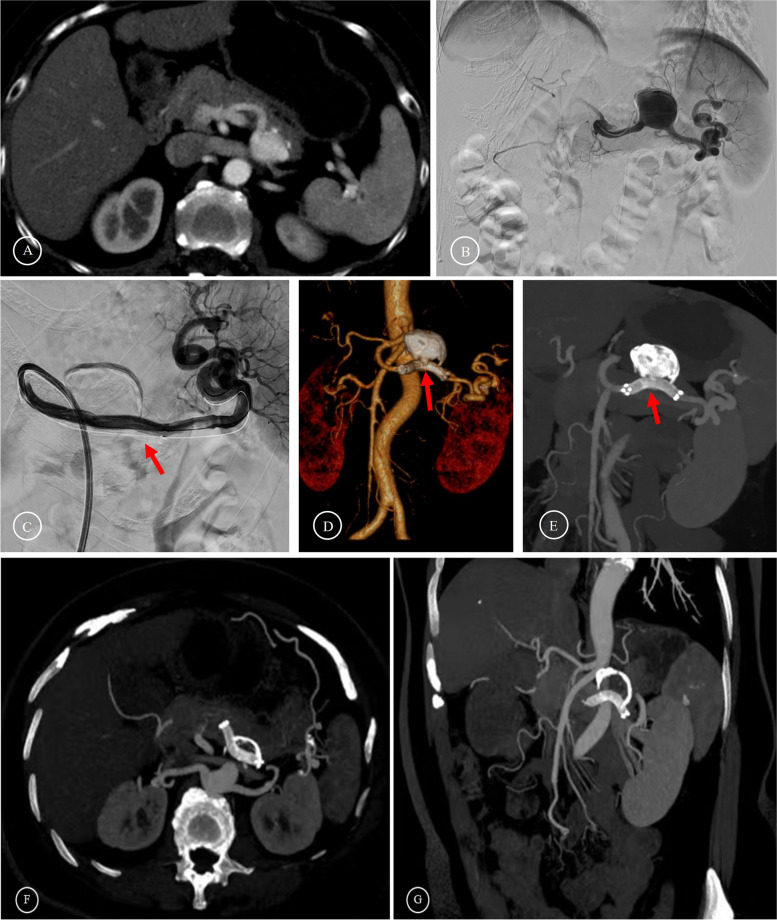
Fig. 4.
Preoperative and postoperative images of stent graft endovascular exclusion A-B Preoperative images showed that the SAA was located at the mid-section of the SA. C The stent graft (Viabahn) was released to isolate the aneurysm. D-E The follow-up two months after the operation showed that the blood flow in the stent graft was unobstructed. (The red arrows indicate the stent graft.) F-G The follow-up three years after the operation showed the calcium wrap around the aneurysm

